Positron Emission Tomography-CT, CT, and MR Imaging Findings of Tumor-Mimicking Organized Hematoma in the Maxillary Sinus: Two Case Reports
- Affiliations
-
- 1Department of Radiology, Chungnam National University Hospital, Chungnam National University School of Medicine, Daejeon, Korea. haneul88@hanmail.net
- 2Department of Pathology, Chungnam National University Hospital, Chungnam National University School of Medicine, Daejeon, Korea.
- KMID: 1443472
- DOI: http://doi.org/10.3348/jksr.2011.65.2.113
Abstract
- Here in, we report two cases of organized hematoma in the maxillary sinus mimicking inverted papilloma. These cases presented as heterogeneously enhancing, expansile masses on computed tomography and magnetic resonance imaging, and also evidenced mild uneven hypermetabolism on positron emission tomography-computed tomography. Organized hematoma should be included in the differential diagnosis of inverted papilloma.
MeSH Terms
Figure
-
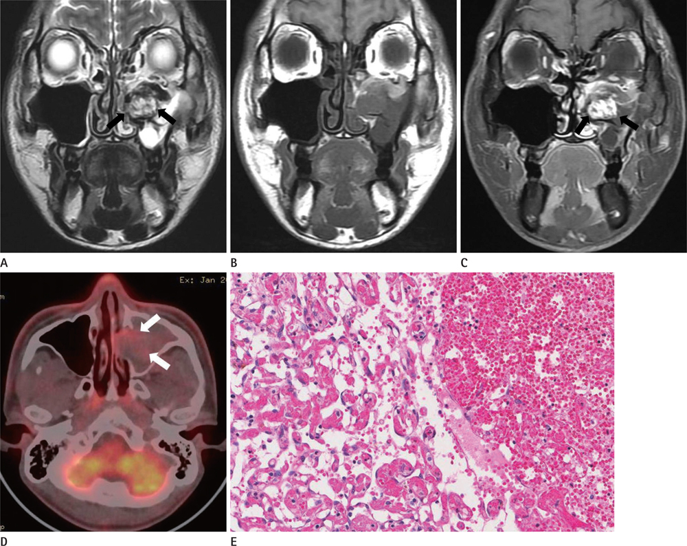
Fig. 1 MR, PET imaging, and histopathologic findings for case 1. A. Coronal T2-weighted fast spin-echo image depicts a heterogeneous high-signal intensity mass (arrow) with a hypointense peripheral rim in the left maxillary sinus. B, C. Coronal T1-weighted spin-echo image and contrast-enhanced T1-weighted spin-echo coronal images show a highly heterogeneously enhancing mass (arrow) in the left maxillary sinus. D. PET-CT axial image shows subtle uneven FDG uptake (maximum SUV = 2.7) in the mass (arrow). E. Hematoxylin-eosin staining (× 100) shows endothelialization of the organized hematoma from case 1. Note.-FDG = fluorodeoxyglucose, PET = positron emission tomography, SUV = standard uptake value

Fig. 2 MR and PET imaging for Case 2. A. Axial T2-weighted fast spin-echo image shows a cauliflower-shaped, heterogeneous hyperintense mass with a dark signal at the peripheral rim. B. PET-CT axial image shows increased FDG uptake (maximum SUV = 4.3) at the peripheral portion of the mass (arrow). C. Hematoxylin-eosin staining (× 100) shows organized thrombus replaced by vascularized fibrotic tissue. Note.-FDG = fluorodeoxyglucose, PET = positron emission tomography, SUV = standard uptake value
Reference
-
1. Lee PK, Wu JK, Ludemann JP. Hemorrhagic pseudotumour of the maxillary sinus. J Otolaryngol. 2004; 33:206–208.2. Lee HK, Smoker WR, Lee BJ, Kim SJ, Cho KJ. Organized hematoma of the maxillary sinus: CT findings. AJR Am J Roentgenol. 2007; 188:W370–W373.3. Yoon TM, Kim JH, Cho YB. Three cases of organized hematoma of the maxillary sinus. Eur Arch Otorhinolaryngol. 2006; 263:823–826.4. Song HM, Jang YJ, Chung YS, Lee BJ. Organizing hematoma of the maxillary sinus. Otolaryngol Head Neck Surg. 2007; 136:616–620.5. Ojiri H, Ujita M, Tada S, Fukuda K. Potentially distinctive features of sinonasal inverted papilloma on MR imaging. AJR Am J Roentgenol. 2000; 175:465–468.6. Ozhan S, Araç M, Isik S, Oznur II, Atilla S, Kemaloglu Y. Pseudotumor of the maxillary sinus in a patient with von Willebrand's disease. AJR Am J Roentgenol. 1996; 166:950–951.7. Kim EY, Kim HJ, Chung SK, Dhong HJ, Kim HY, Yim YJ, et al. Sinonasal organized hematoma: CT and MR imaging findings. AJNR Am J Neuroradiol. 2008; 29:1204–1208.8. Tabaee A, Kacker A. Hematoma of the maxillary sinus presenting as a mass--a case report and review of literature. Int J Pediatr Otorhinolaryngol. 2002; 65:153–157.9. Lee BJ, Park HJ, Heo SC. Organized hematoma of the maxillary sinus. Acta Otolaryngol. 2003; 123:869–872.10. Jeon TY, Kim HJ, Choi JY, Lee IH, Kim ST, Jeon P, et al. 18F-FDG PET/CT findings of sinonasal inverted papilloma with or without coexistent malignancy: comparison with MR imaging findings in eight patients. Neuroradiology. 2009; 51:265–271.
- Full Text Links
-
- Actions
-
Cited
- CITED
-
- Close
- Share
-
- Similar articles
-
- Organized Hematoma Presenting with Periorbital Swelling: A Case Report and Review of Literatures
- Organized Hematoma of the Maxillary Sinus: Surgical Excision by Midfacial Degloving Approach
- A Case of Organized Hematoma after Coblation Assisted Turbinoplasty
- Maxillary Sinus Mucocele Secondary to Organized Hematoma
- Oral cancer diagnosed using PET/CT: A case report
