A Case of Bilateral Trochlear Nerve Palsy Following Cisternography
- Affiliations
-
- 1Department of Ophthalmology, Catholic University of Daegu School of Medicine, Daegu, Korea. kimsy@cu.ac.kr
Abstract
- PURPOSE
To report a case of bilateral trochlear nerve palsy following cisternography.
CASE SUMMARY
A 43-year-old male with intermittent watery rhinorrhea persisting for 3 months visited the neurosurgery department of our institute. His past medical history included removal of a pituitary adenoma 22 years prior to presentation. Cerebrospinal fluid leakage was suspected and cisternography was performed. The patient was referred to our ophthalmology department for diplopia 3 days after the cisternography. An alternate prism cover test showed 5 prism diopter (PD) right hypertrophia in the primary position, and underaction of bilateral superior oblique muscles and overaction of the left inferior oblique muscle. A positive Bielschowsky test with the head tilted to either side was observed and excyclotorsion was 9degrees on the double Maddox rod test. The patient was diagnosed with bilateral trochlear nerve palsy. After 2 years of follow-up, diplopia persisted and recession of the bilateral inferior oblique muscles was performed. After the surgery, diplopia disappeared, the fundus photography showed no excyclotorsion, and the double Maddox rod test indicated 3degrees of excyclotorsion.
CONCLUSIONS
Cisternography should be carefully performed due to the possibility of bilateral trochlear nerve palsy, an extremely rare but possible occurrence following the procedure.
MeSH Terms
Figure
-
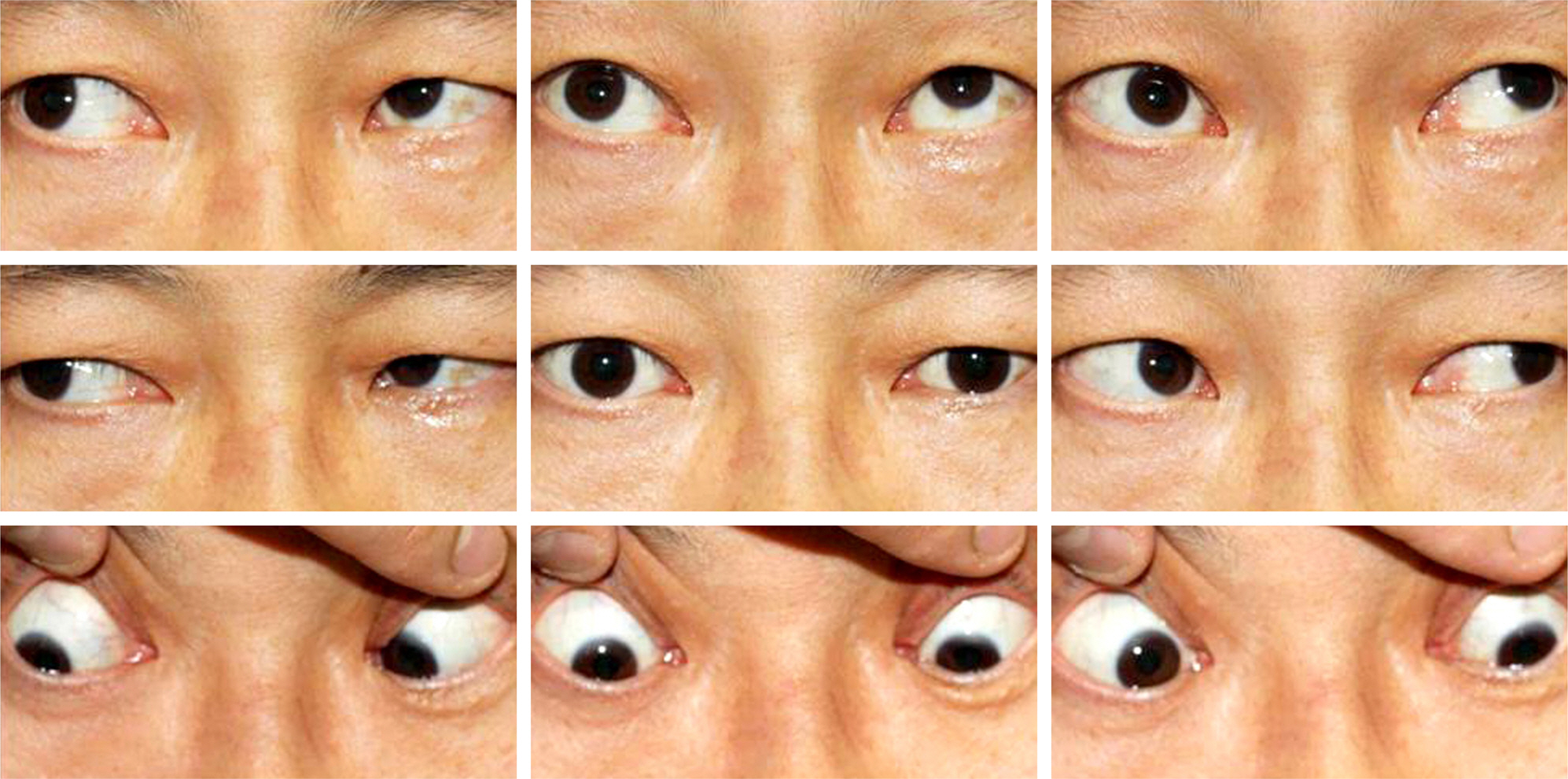
Figure 1. Nine cardinal photographs at the first visit showing underaction of bilateral superior oblique muscles and overaction of the left inferior oblique muscle.
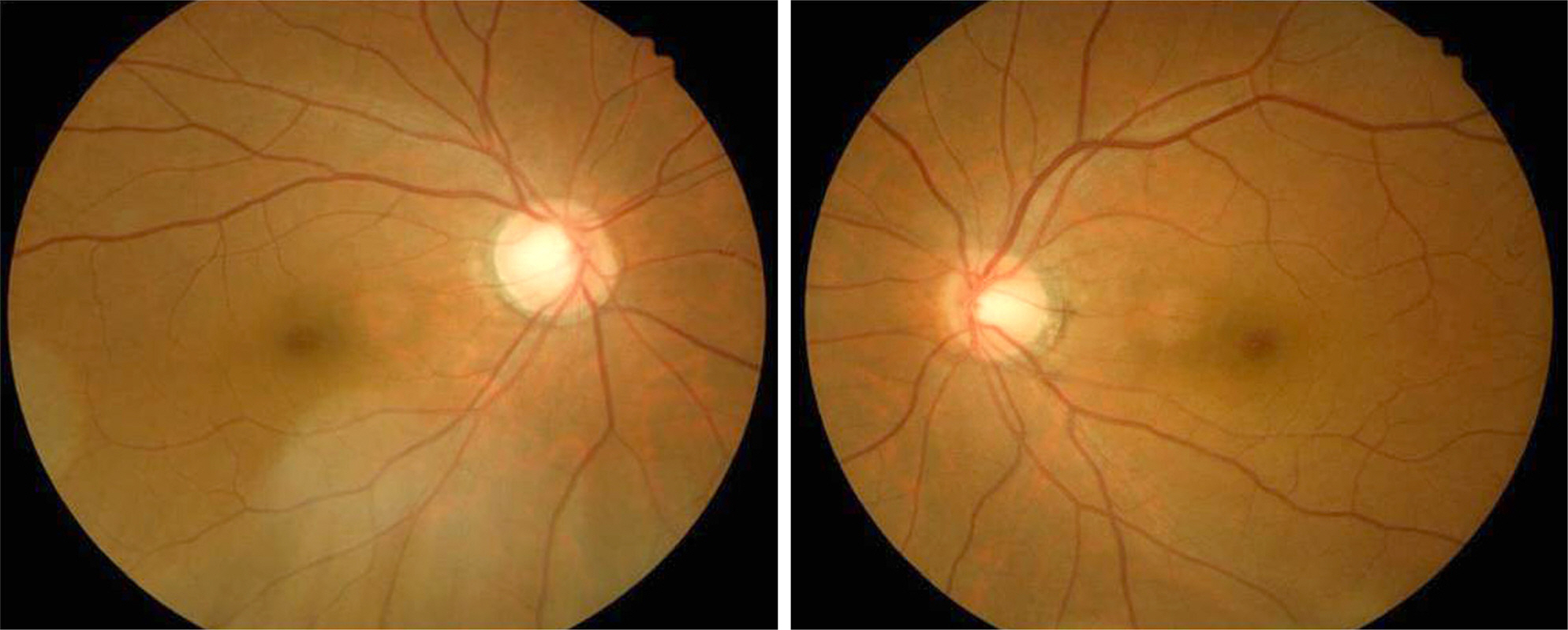
Figure 2. Fundus photographs at the first visit showing excyclotorsion in the right eye.
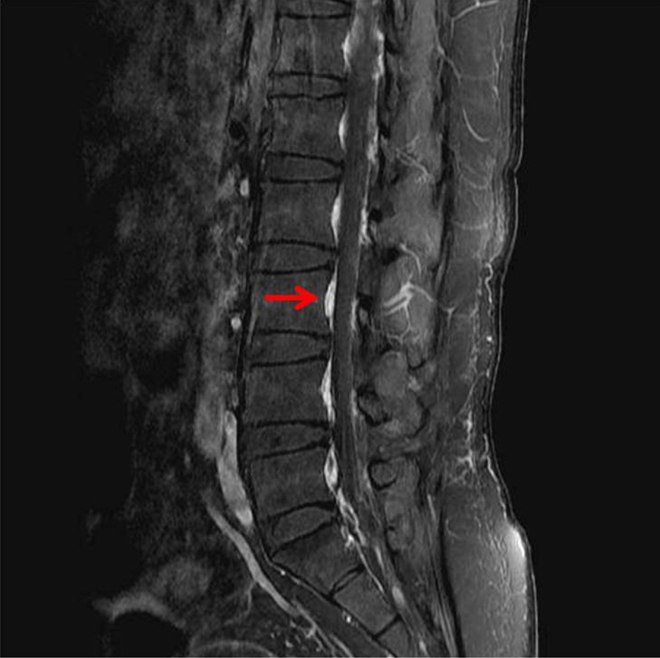
Figure 3. Lumbar spine MRI with postcontrast fat-suppressed T1-weighted sagittal image showing increased epidural enhancement (arrow).
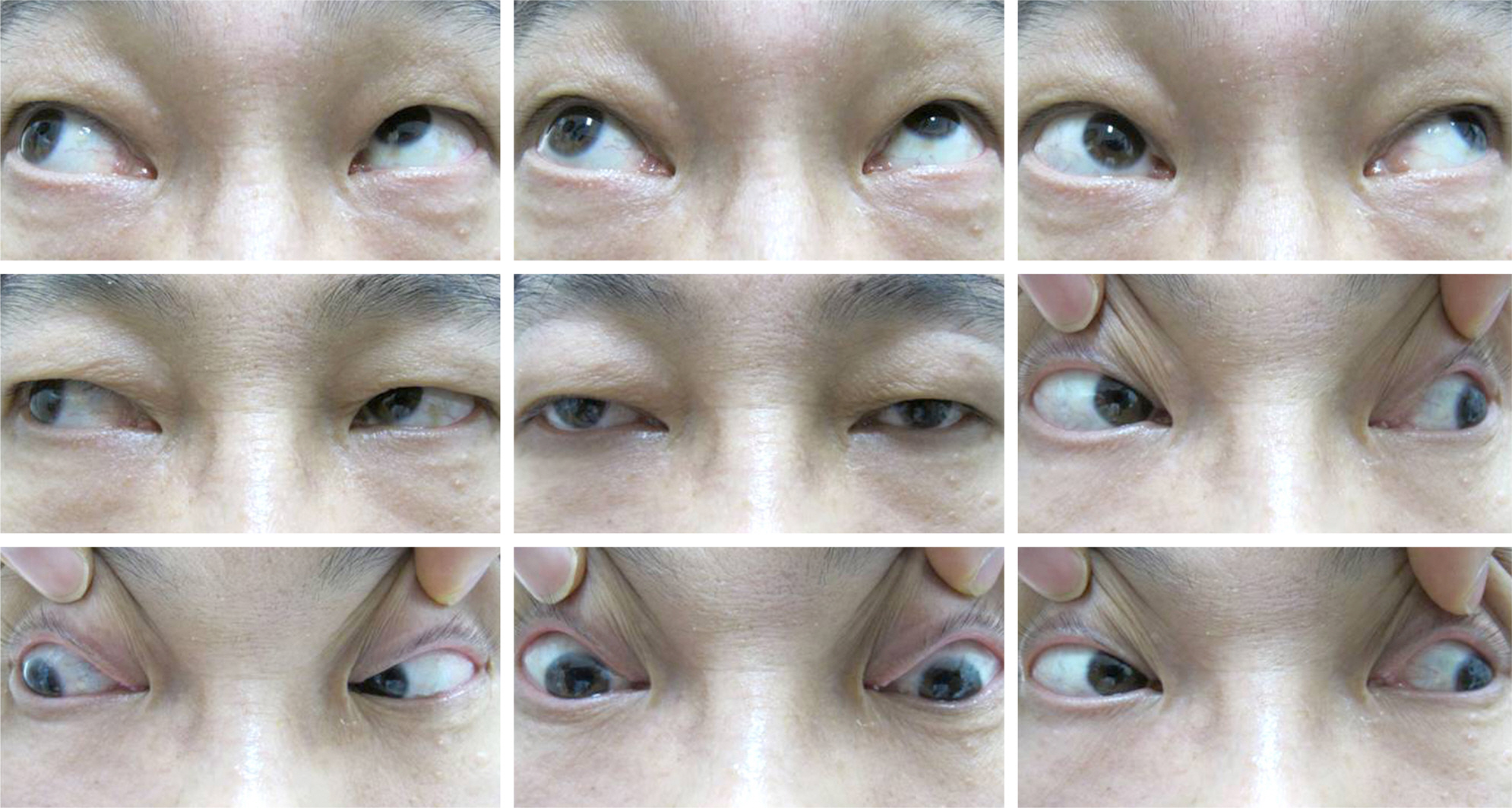
Figure 4. Preoperative nine cardinal photographs showing underaction of bilateral superior oblique muscles and marked overaction of the left inferior oblique muscle.

Figure 5. Hess screen test chart of the patient with bilateral trochlear nerve palsy.
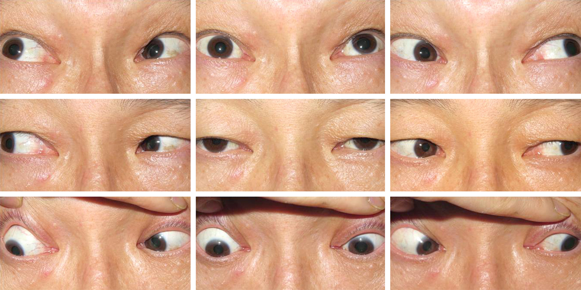
Figure 6. Nine cardinal photographs after recession of the bilateral inferior oblique muscles presenting no overaction of bilateral inferior oblique muscles and mild underaction of the left superior oblique muscle.
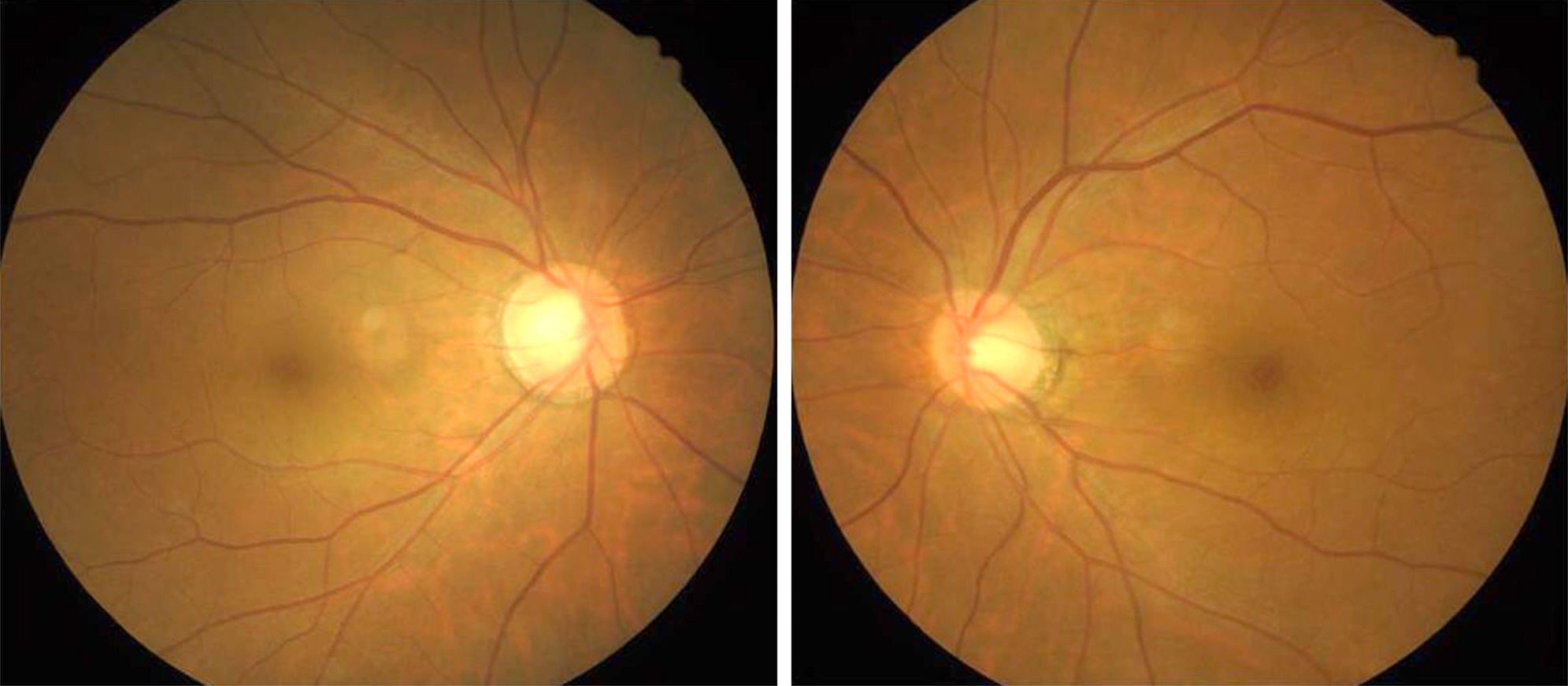
Figure 7. Fundus photographs after weakening of bilateral inferior oblique muscles showing no excyclotorsion in both eyes.
Reference
-
References
1. King RA, Calhoun JH.Fourth cranial nerve palsy following spinal anesthesia. A case report. J Clin Neuroophthalmol. 1987; 7:20–2.2. Thorsén G.Neurological complications after spinal anaesthesia: and results from 2493 follow-up cases. Acta Chirg Scand. 1947; 95:75–94.3. Follens I, Godts D, Evens PA, Tassignon MJ.Combined fourth and sixth cranial nerve palsy after lumbar puncture: a rare complication. A case report. Bull Soc Belge Ophtalmol. 2001(281):29–33.4. Mansour AM, Reinecke RD.Central trochlear palsy. Surv Ophthalmol. 1986; 30:279–97.
Article5. Sakurai K, Nishio M, Yamada K. . Comparison of the radio-isotope cisternography findings of spontaneous intracranial hypo-tension and iatrogenic cerebrospinal fluid leakage focusing on chronological changes. Cephalalgia. 2012; 32:1131–9.
Article6. Levine MC, Jayabalan V.Complications of isotope cisternography. Ann Neurol. 1977; 1:172–6.
Article7. Guy G, Comoy J, Chevet D. . [Aseptic meningitis after isotope cisternography. 2 Cases]. Neurochirurgie. 1976; 22:631–8.8. Ponto JA.Special safety considerations in preparation of techne-tium Tc-99m DTPA for cerebrospinal fluid-related imaging procedures. J Am Pharm Assoc (2003). 2008; 48:413–6.
Article9. Park BS, Park J, Koh SH. . Conus medullaris syndrome as a complication of radioisotope cisternography. Can J Neurol Sci. 2012; 39:347–51.
Article10. Amoozegar F, Guglielmin D, Hu W. . Spontaneous intracranial hypotension: recommendations for management. Can J Neurol Sci. 2013; 40:144–57.
Article11. Zada G, Solomon TC, Giannotta SL.A review of ocular manifes-tations in intracranial hypotension. Neurosurg Focus. 2007; 23:E8.
Article
- Full Text Links
-
- Actions
-
Cited
- CITED
-
- Close
- Share
-
- Similar articles
-
- A Case of Herpes Zoster Ophthalmicus with Isolated Trochlear Nerve Involvement
- A Case with Bilateral Dural Arteriovenous Fistulae Manifesting as Sequential Trochlear, Oculomotor Nerve Palsies
- A Clinical Study of Diplopia Due to Neurologic Disorder
- Combined Facial and Contralateral Trochlear Nerve Palsy in a Patient with Diabetes Mellitus
- Transient Isolated Trochlear Nerve Palsy Associated with Rathke's Cleft Cyst
