J Korean Ophthalmol Soc.
2013 Oct;54(10):1599-1604.
Malignant Solitary Fibrous Tumor of the Orbit
- Affiliations
-
- 1Department of Ophthalmology, Samsung Medical Center, Sungkyunkwan University School of Medicine, Seoul, Korea. ydkimoph@skku.edu
Abstract
- PURPOSE
Solitary fibrous tumor (SFT) is a rare tumor of the pleura, mediastinum, pericardium and other organs affecting predominantly middle-aged patients. SFT arising in the orbit is extremely rare, and its malignant form is even rarer. The authors herein describe a case of malignant solitary fibrous tumor of the orbit.
CASE SUMMARY
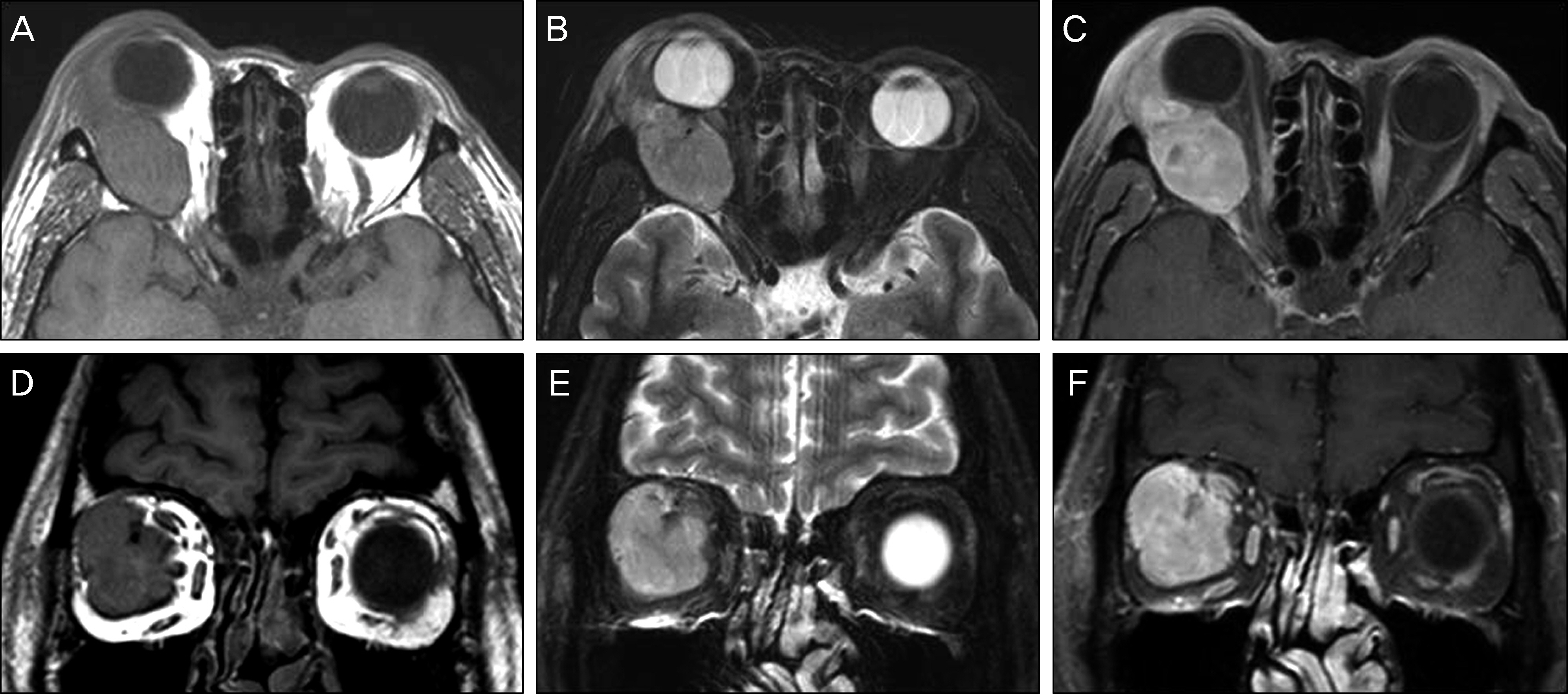
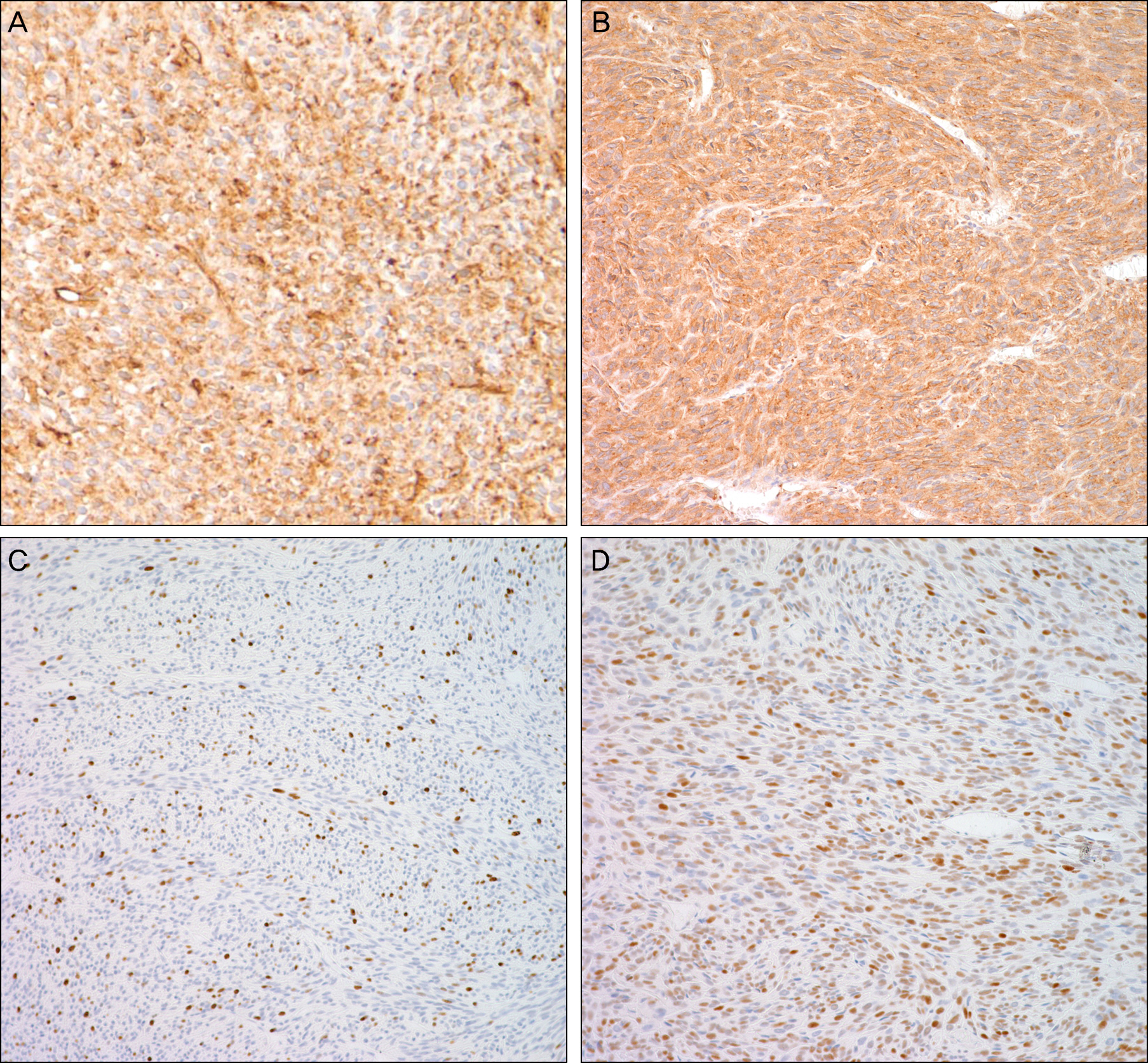
A 67-year-old male presented with a 4-month history of right proptosis. On ophthalmologic examination, 9-mm proptosis was observed in the right eye, and extraocular movements were limited in all directions of gaze. CT scan and MR imaging showed a lobulated, well-enhancing mass adjacent to the lateral rectus muscle in the superolateral retrobulbar space. Excisional biopsy through a lateral orbitotomy was performed. Histopathological examination showed proliferation of spindle cells with a fascicular pattern interspersed with bands of collagen, increased cellularity, cellular pleomorphism, hemorrhage, necrosis and high mitotic activity. Immunohistochemical staining revealed diffuse positivity for CD34, CD99, Ki67 and p53, and malignant SFT was diagnosed.
CONCLUSIONS
Malignant SFT should be considered in the differential diagnosis of acutely progressing unilateral proptosis.
Keyword
MeSH Terms
Figure
Reference
-
References
1. Klemperer P, Rabin CB. Primary neoplasm of the pleura: a report of five cases. Arch Pathol. 1931; 11:385–412.2. Bernardini FP, de Conciliis C, Schneider S. . Solitary fibrous tumor of the orbit. Is it rare? Report of a case series and review of the literature. Ophthalmology. 2003; 110:1442–8.3. Dorfman DM, To K, Dickersin GR. . Solitary fibrous tumor of the orbit. Am J Surg Pathol. 1994; 18:281–7.
Article4. Cho NH, Kie JH, Yang WI, Jung WH. Solitary fibrous tumor with an unusual adenofibromatous feature in the lacrimal gland. Histopathology. 1998; 33:289–90.5. Woo KI, Suh YL, Kim YD. Solitary fibrous tumor of the lacrimal sac. Ophthal Plast Reconstr Surg. 1999; 15:450–3.
Article6. Kim EK, Paik JS, Yang SW. A case of solitary fibrous tumor of orbit. J Korean Ophthalmol Soc. 2010; 51:881–4.
Article7. Mascarenhas L, Lopes M, Duarte AM. . Histologically malignant solitary fibrous tumor of the orbit. Neurochirurgie. 2006; 52:415–8.
Article8. Girnita L, Sahlin S, Orrego A. . Malignant solitary fibrous tumour of the orbit. Acta Ophthalmol. 2009; 87:464–7.
Article9. Manousaridis K, Stropahl G, Guthoff RF. Recurrent malignantsolitary fibrous tumor of the orbit. Ophthalmologe. 2011; 108:260–4.10. Brunnemann RB, Ro JY, Ordonez NG. . Extrapleural solitary fibrous tumour: a clinicopathologic study of 24 cases. Mod Pathol. 1999; 12:1034–42.11. Churg A, Cagle PT, Roggli VL. Solitary fibrous tumor. Tumors of the serosal membranes. 4th ed.Washington DC: American Registry of Pathology Press;2006. p. 299.12. Ohtsuka K, Hashimoto M, Suzuki Y. A review of 244 orbital tumours in Japanese patients during a 21-year period: origins and locations. Jpn J Ophthalmol. 2005; 49:49–55.13. Shields JA, Shields CL, Scartozzi R. Survey of 1264 patients with orbital tumours and simulating lesions: the 2002 Montgomery Lecture, Part 1. Ophthalmology. 2004; 111:997–1008.14. Saeed P, Rootman J, Nugent RA. . Optic nerve sheath meningiomas. Ophthalmology. 2003; 110:2019–30.
Article15. Westra WH, Gerald WL, Rosai J. Solitary fibrous tumor. Consistent CD34 immunoreactivity and occurrence in the orbit. Am J Surg Pathol. 1994; 18:992–8.
Article16. England DM, Hochholzer L, McCarthy MJ. Localized benign and malignant fibrous tumors of the pleura: a clinicopathologic review of223 cases. Am J Surg Pathol. 1989; 13:640–58.17. Cassarino DS, Auerbach A, Rushing EJ. Widely invasive solitary fibrous tumor of the sphenoid sinus, cavernous sinus, and pituitary fossa. Ann Diagn Pathol. 2003; 7:169–73.
Article18. Suster S, Fisher C, Moran CA. . Expression of bcl-2 oncoprotein in benign and malignant spindle cell tumors of soft tissue, skin, serosal surfaces, and gastrointestinal tract. Am J Surg Pathol. 1998; 22:863–72.
Article19. Guillou L, Fletcher JA, Fletcher CDM, Mandahl N. Extrapleural solitary fibrous tumor and hemangiopericytoma. Fletcher CDM, Unni KK, Mertens F Lyon, editors. Pathology and genetics of tumours of soft tissues and bone. France: IARC Press;2002. p. 86.20. Sun Y, Naito Z, Ishiwata T. . Basic FGF and Ki-67 proteins useful for immunohistological diagnostic evaluations in malignant solitary fibrous tumor. Pathol Int. 2003; 53:284–90.
Article21. Yokoi T, Tsuzuki T, Yatabe Y. . Solitary fibrous tumour: significance of p53 and CD34 immunoreactivity in its malignant transformation. Histopathology. 1998; 32:423–32.
Article22. Furusato E, Valenzuela IA, Fanburg-Smith JC. . Orbital solitary fibrous tumor: encompassing terminology for hemangiopericytoma, giant cell angiofibroma, and fibrous histiocytoma of the orbit: reappraisal of 41 cases. Hum Pathol. 2011; 42:120–8.
Article23. Gigantelli JW, Kincaid MC, Soparkar CN. . Orbital solitary fibrous tumor: radiographic and histopathologic correlations. Ophthal Plast Reconstr Surg. 2001; 17:207–14.
Article24. Romer M, Bode B, Schuknecht B. . Solitary fibrous tumor of the orbit–two cases and a review of the literature. Eur Arch Otorhinolaryngol. 2005; 262:81–8.
Article25. Yang BT, Wang YZ, Dong JY. . MRI study of solitary fibrous tumor in the orbit. AJR Am J Roentgenol. 2012; 199:W506–11.
Article26. Dalley RW. Fibrous histiocytoma and fibrous tissue tumors of the orbit. Radiol Clin North Am. 1999; 37:185–94.
Article27. Meyer D, Riley F. Solitary fibrous tumor of the orbit: a clin- icopathologic entity that warrants both a heightened awareness and an atraumatic surgical removal technique. Orbit. 2006; 25:45–50.28. Alexandrakis G, Johnson TE. Recurrent orbital solitary fibrous tumour in a 14-year-old girl. Am J Ophthalmol. 2000; 130:373–6.29. Hayashi S, Kurihara H, Hirato J, Sasaki T. Solitary fibrous tumor of the orbit with extraorbital extension: case report. Neurosurgery. 2001; 49:1241–5.
Article


