J Korean Ophthalmol Soc.
2012 Oct;53(10):1403-1411.
Changes in Tear Film, Cornea and Ocular Surface According to the Duration of Soft Contact Lens Wear
- Affiliations
-
- 1Cheil Eye Hospital, Daegu, Korea. eyepark9@dreamwiz.com
Abstract
- PURPOSE
The present study evaluated the changes in tear film, cornea and ocular surface according to the duration of soft contact lens wear.
METHODS
A total 65 patients with 130 eyes were enrolled the present study, and were divided into 4 grous. The control group (group A) was composed of 32 eyes of 16 patients who had not worn soft contact lenses, group B, (34 eyes of 17 patients), had worn soft contact lenses less than 5 years, group C, (32 eyes of 16 patients), had worn soft contact lenses for 5 to 10 years and group D, (32 eyes of 16 patients), had worn soft contact lenses for more than 10 years. The tear break-up time (BUT), Schirmer's test, corneal sensitivity test, central corneal thickness, ocular surface disease index (OSDI), corneal fluorescein staining score, specular microscopy and conjunctival impression cytology were analyzed. The results were compared between the control group and the soft contact lens groups.
RESULTS
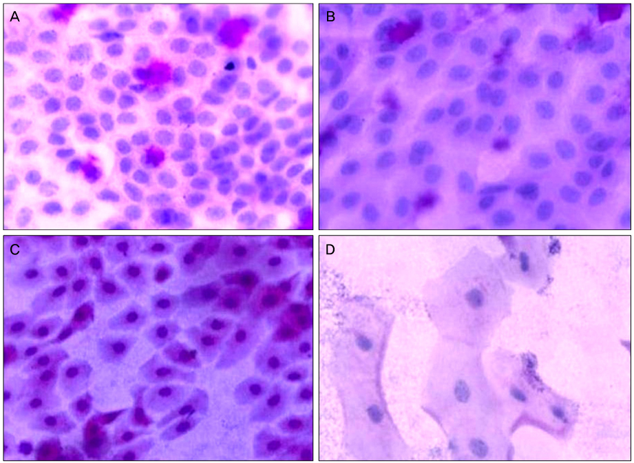
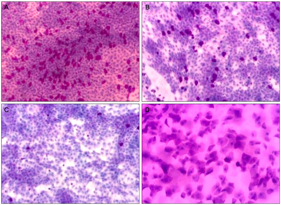
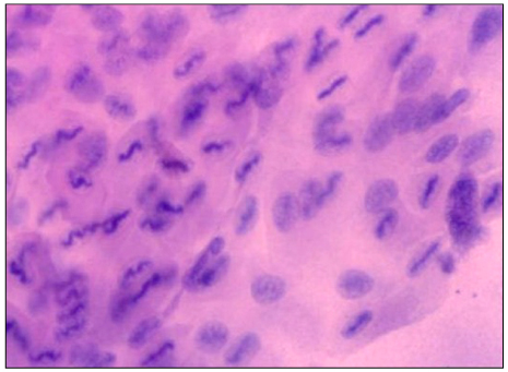
In group B, BUT significantly decreased, but corneal fluorescein staining score and squamous metaplasia significantly increased (p < 0.05). In group C, OSDI and snake-like chromatin pattern significantly increased, but corneal thickness and goblet cell density significantly decreased (p < 0.05). In group D, the coefficient of variation for endothelium significantly increased, but corneal sensitivity and hexagonality significantly decreased (p < 0.05).
CONCLUSIONS
Duration of soft contact lens wear influences changes in tear film, cornea and ocular surface.
Keyword
MeSH Terms
Figure
Reference
-
1. Korean Contact Lens Society. Contact Lens: Principles and Practice. 2007. Vol. 1:2nd ed. Seoul: Nae-Oi Publishing & Printing;187–202.2. Gürdal C, Aydin S, Kirimlioğlu H, et al. Effects of extended-wear soft contact lenses on the ocular surface and central corneal thickness. Ophthalmologica. 2003. 217:329–336.3. Hori Y, Argüeso P, Spurr-Michaud S, Gipson IK. Mucins and contact lens wear. Cornea. 2006. 25:176–181.4. Glasson MJ, Stapleton F, Keay L, et al. Differences in clinical parameters and tear film of tolerant and intolerant contact lens wearers. Invest Ophthalmol Vis Sci. 2003. 44:5116–5124.5. Shin YJ, Moon JW, Wee WR. Corneal neovascularization and corneal hypesthesia as contact lens Complications. J Korean Ophthalmol Soc. 2006. 47:25–30.6. Murphy PJ, Patel S, Marshall J. The effect of long-term, daily contact lens wear on corneal sensitivity. Cornea. 2001. 20:264–269.7. Brennan NA, Bruce AS. Esthesiometry as an indicator of corneal health. Optom Vis Sci. 1991. 68:699–702.8. Liu Z, Pflugfelder SC. The effects of long-term contact lens wear on corneal thickness, curvature, and surface regularity. Ophthalmology. 2000. 107:105–111.9. Braun DA, Anderson Penno EE. Effect of contact lens wear on central corneal thickness measurements. J Cataract Refract Surg. 2003. 29:1319–1322.10. Holden BA, Sweeney DF, Vannas A, et al. Effects of long-term extended contact lens wear on the human cornea. Invest Ophthalmol Vis Sci. 1985. 26:1489–1501.11. Park YJ, Lee GJ, Park JJ, et al. The long-term effects of soft contact lens wear on corneal thickness, curvature and endothelium. J Korean Ophthalmol Soc. 2005. 46:945–953.12. Doh HJ, Joo CK. Effect of aging and soft contact lens wearing on the change of corneal endothelial cells. J Korean Ophthalmol Soc. 1999. 40:330–337.13. Setälä K, Vasara K, Vesti E, Ruusuvaara P. Effects of long-term contact lens wear on the corneal endothelium. Acta Ophthalmol Scand. 1998. 76:299–303.14. Egbert PR, Lauber S, Maurice DM. A simple conjunctival biopsy. Am J Ophthalmol. 1977. 84:798–801.15. Cakmak SS, Unlü MK, Karaca C, et al. Effects of soft contact lenses on conjunctival surface. Eye Contact Lens. 2003. 29:230–233.16. Anshu , Munshi MM, Sathe V, Ganar A. Conjunctival impression cytology in contact lens wearers. Cytopathology. 2001. 12:314–320.17. Adar S, Kanpolat A, Sürücü S, Ucakhan OO. Conjunctival impression cytology in patients wearing contact lenses. Cornea. 1997. 16:289–294.18. Tomatir DK, Erda N, Gürlü VP. Effects of different contact lens materials and contact lens-wearing periods on conjunctival cytology in asymptomatic contact lens wearers. Eye Contact Lens. 2008. 34:166–168.19. Knop E, Brewitt H. Conjunctival cytology in asymptomatic wearers of soft contact lenses. Graefes Arch Clin Exp Ophthalmol. 1992. 230:340–347.20. Bron AJ, Evans VE, Smith JA. Grading of corneal and conjunctival staining in the context of other dry eye tests. Cornea. 2003. 22:640–650.21. Nelson JD. Impression cytology. Cornea. 1988. 7:71–81.22. Lee DK, Choi SK, Song KY. Clinical survey of corneal complications associated with contact lens wear. J Korean Ophthalmol Soc. 1994. 35:895–901.23. Park M, Kwak C, Tchah H. Experimental 24 hour corneal swelling by extended wear contact lenses. J Korean Ophthalmol Soc. 1991. 32:149–153.24. Foulks GN. What is dry eye and what does it mean to the contact lens wearer? Eye Contact Lens. 2003. 29:S96–S100. S115–S118. S192–S194.25. Kangas TA, Edelhauser HF, Twining SS, O'Brien WJ. Loss of stromal glycosaminoglycans during corneal edema. Invest Ophthalmol Vis Sci. 1990. 31:1994–2002.26. Martin DK. Osmolality of the tear fluid in the contralateral eye during monocular contact lens wear. Acta Ophthalmol (Copenh). 1987. 65:551–555.27. Helena MC, Baerveldt F, Kim WJ, Wilson SE. Keratocyte apoptosis after corneal surgery. Invest Ophthalmol Vis Sci. 1998. 39:276–283.28. Stefansson E, Wolbarsht ML, Landers MB 3rd. The corneal contact lens and aqueous humor hypoxia in cats. Invest Ophthalmol Vis Sci. 1983. 24:1052–1054.29. Knop E, Reale E. Fine structure and significance of snakelike chromatin in conjunctival epithelial cells. Invest Ophthalmol Vis Sci. 1994. 35:711–719.30. Marner K. 'Snake-like' appearance of nuclear chromatin in conjunctival epithelial cells from patients with keratoconjunctivitis sicca. Acta Ophthalmol (Copenh). 1980. 58:849–853.31. Knop E, Brewitt H. Induction of conjunctival epithelial alterations by contact lens wearing. A prospective study. Ger J Ophthalmol. 1992. 1:125–134.32. Maudgil SS, Khurana AK, Singh M, et al. Tear film flow and stability in normal Indian subjects. Indian J Ophthalmol. 1989. 37:182–183.33. Ozdemir M, Temizdemir H. Age- and gender-related tear function changes in normal Population. Eye (Lond). 2010. 24:79–83.34. Kim SD, Kim JK, Han HK. Normal conjunctival goblet cell density in Korean measured by impression cytology. J Korean Ophthalmol Soc. 1992. 33:427–435.
- Full Text Links
- Actions
-
Cited
- CITED
-
- Close
- Share
- Similar articles
-
- Clinical Use of Mini-Scleral Contact Lens in Ocular Surface Diseases
- The Study of Tear Film Break Up Time(BUT) in Soft Contact Lens Wearer
- The Lysozymal Concentration in Tear Film of Contact Lens Us ers
- The Effects of Cheap Tinted Contact Lenses on Corneal Swelling and Ocular Surface Inflammation
- The effect of Contact Lens Wear on Tear Secretion


