A Case of Localized Merkel Cell Carcinoma on the Upper Eyelid
- Affiliations
-
- 1Sungmo Eye Hospital, Busan, Korea. rhoahn@yahoo.co.kr
Abstract
- PURPOSE
To report a case of Merkel cell carcinoma on the left upper eyelid without metastasis and its immunohistochemical features. The carcinoma was successfully treated with excisional surgery and prophylactic radiation therapy.
CASE SUMMARY
A 76-year-old woman presented to the hospital complaining of a 0.6 x 0.9-cm-sized painless and purplish-red colored mass that had grown rapidly on her left upper eyelid margin over the previous two months. An excisional biopsy was performed. On immunohistochemical examination of the lesion, the tumor cells expressed immunoreactivity for synaptophysin and were negative for LAC and cytokeratin, confirming the diagnosis of Merkel cell carcinoma. Additional surgery was performed because the surgical margins were positive. No other primary or metastatic lesions were found. The patient was treated with local prophylactic irradiation and remained disease-free at her 10-month follow-up visit.
Keyword
MeSH Terms
Figure
-
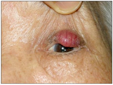
Figure 1 The lesion of the eyelid was nodular, nontender, and had relatively well-defined margins, measuring about 0.9 × 1.3 cm. It was fixed to tarsus and purplish-red in color. The overlying skin was intact with telangiectasia.

Figure 2 (A) The tumor cells show uniform size with a round to oval nucleus and scanty cytoplasm. Arrow indicates vascular invasion (H&E stain, ×200). (B) The blue tumor cells infiltrate into the hair follicle. Arrow indicates hair follicle (H&E stain, ×100).

Figure 3 Immunohistochemistry for synaptophysin indicating neuroendodermal origin, show brown positive staining on the tumors (×100).

Figure 4 Immunohistochemistry for leukocyte common antigen shows negative (×100).

Figure 5 Tumor cells are negative for cytokeratin (×100).

Figure 6 Eight-month postoperative view. Functionally and cosmetically satisfactory results were obtained without any recurrence.
Reference
-
1. Toker C. Trabecular carcinoma of the skin. Arch Dermatol. 1972. 105:107–110.2. Barrett RV, Meyer DR. Eyelid and periocular cutaneous merkel cell carcinoma (aka. Neuroendocrine or trabecular carcinoma). Int Ophthalmol Clin. 2009. 49:63–75.3. Ikawa F, Kiya K, Uozumi T, et al. Brain metastasis of Merkel cell carcinoma. Case report and review of the literature. Neurosurg Rev. 1999. 22:54–57.4. Huang GS, Chang WC, Lee HS, et al. Merkel cell carcinoma arising from the subcutaneous fat of the arm with intact skin. Dermatol Surg. 2005. 31:717–719.5. Raaf JH, Urmacher C, Knapper WK, et al. Trabecular (Merkel cell) carcinoma of the skin. Treatment of primary, recurrent, and metastatic disease. Cancer. 1986. 57:178–182.6. Mamalis N, Medlock RD, Holds JB, et al. Merkel cell tumor of the eyelid: a review and report of an unusual case. Ophthalmic Surg. 1989. 20:410–414.7. Turk T, Orlic ZC, Smoljan I, et al. Spontaneous regression of Merkel cell carcinoma in a patient with chronic lymphocytic leukemia: a case report. J Med Case Rep. 2009. 3:7270.8. Junquera L, Torre A, Vicente JC, et al. Complete spontaneous regression of Merkel cell carcinoma. Ann Otol Rhinol Laryngol. 2005. 114:376–380.9. Nicoletti AG, Matayoshi S, Santo RM, Ferreira VR. Eyelid Merkel cell carcinoma: report of three cases. Ophthal Plast Reconstr Surg. 2004. 20:117–121.10. Tanahashi J, Kashima K, Daa T, et al. Merkel cell carcinoma co-existent with sebaceous carcinoma of the eyelid. J Cutan Pathol. 2009. 36:983–986.11. Soltau JB, Smith ME, Custer PL. Merkel cell carcinoma of the eyelid. Am J Ophthalmol. 1996. 121:331–332.12. Colombo F, Holbach LM, Junemann AG, et al. Merkel cell carcinoma: clinicopathologic correlation, management, and follow-up in five patients. Ophthal Plast Reconstr Surg. 2000. 16:453–458.13. Rawlings NG, Brownstein S, Jordan DR. Merkel cell carcinoma masquerading as a chalazion. Can J Ophthalmol. 2007. 42:469–470.14. Thakur S, Chalioulias K, Hayes M, While A. Bilateral primary Merkel cell carcinoma of the upper lid misdiagnosed as Basal cell carcinoma. Orbit. 2008. 27:139–141.15. Peters GB 3rd, Meyer DR, Shields JA, et al. Management and prognosis of Merkel cell carcinoma of the eyelid. Ophthalmology. 2001. 108:1575–1579.16. Heath M, Jaimes N, Lemos B, et al. Clinical characteristics of Merkel cell carcinoma at diagnosis in 195 patients: the AEIOU features. J Am Acad Dermatol. 2008. 58:375–381.17. Liao PB. Merkel cell carcinoma. Dermatol Ther. 2008. 21:447–451.18. Nicoletti AG, Matayoshi S, Santo RM, Ferreira VR. Eyelid Merkel cell carcinoma: report of three cases. Ophthal Plast Reconstr Surg. 2004. 20:117–121.19. Peters GB 3rd, Meyer DR, Shields JA, et al. Management and prognosis of Merkel cell carcinoma of the eyelid. Ophthalmology. 2001. 108:1575–1579.20. Kivela T, Tarkkanen A. The Merkel cell and associated neoplasms in the eyelids and periocular region. Surv Ophthalmol. 1990. 35:171–187.21. Andea AA, Coit DG, Amin B, Busam KJ. Merkel cell carcinoma: histologic features and prognosis. Cancer. 2008. 113:2549–2558.22. Gandhi RK, Rosenberg AS, Somach SC. Merkel cell polyomavirus: an update. J Cutan Pathol. 2009. 36:1327–1329.23. Asioli S, Righi A, Volante M, et al. p63 expression as a new prognostic marker in Merkel cell carcinoma. Cancer. 2007. 110:640–647.24. Marks S, Radin DR, Chandrasoma P. Merkel cell carcinoma. J Comput Tomogr. 1987. 11:291–293.25. Hanly AJ, Elgart GW, Jorda M, et al. Analysis of thyroid transcription factor-1 and cytokeratin 20 separates merkel cell carcinoma from small cell carcinoma of lung. J Cutan Pathol. 2000. 27:118–120.26. Llombart B, Monteagudo C, López-Guerrero JA, et al. Clinicopathological and immunohistochemical analysis of 20 cases of Merkel cell carcinoma in search of prognostic markers. Histopathology. 2005. 46:622–634.27. Allen PJ, Bowne WB, Jaques DP, et al. Merkel cell carcinoma: prognosis and treatment of patients from a single institution. J Clin Oncol. 2005. 23:2300–2309.28. Rubsamen PE, Tanenbaum M, Grove AS, et al. Merkel cell carcinoma of the eyelid and periocular tissues. Am J Ophthalmol. 1992. 113:674–680.29. Hodgson NC. Merkel cell carcinoma: changing incidence trends. J Surg Oncol. 2005. 89:1–4.30. Allen PJ, Bowne WB, Jaques DP, et al. Merkel cell carcinoma: prognosis and treatment of patients from a single institution. J Clin Oncol. 2005. 23:2300–2309.31. Swann MH, Yoon J. Merkel cell carcinoma. Semin Oncol. 2007. 34:51–56.32. Hitchcock CL, Bland KI, Laney RG 3rd, et al. Neuroendocrine (Merkel cell) carcinoma of the skin. Its natural history, diagnosis, and treatment. Ann Surg. 1988. 207:201–207.33. Mott RT, Smoller BR, Morgan MB. Merkel cell carcinoma: a clinicopathologic study with prognostic implications. J Cutan Pathol. 2004. 31:217–223.34. Gillenwater AM, Hessel AC, Morrison WH, et al. Merkel cell carcinoma of the head and neck: effect of surgical excision and radiation on recurrence and survival. Arch Otolaryngol Head Neck Surg. 2001. 127:149–154.35. Jang JW, Kim TH, Kim HY, Lee SY. Two cases of Merkel cell carcinoma of eyelid and neck. J Korean Ophthalmol Soc. 2000. 41:251–257.36. Bichakjian CK, Lowe L, Lao CD, et al. Merkel cell carcinoma: critical review with guidelines for multidisciplinary management. Cancer. 2007. 110:1–12.
- Full Text Links
-
- Actions
-
Cited
- CITED
-
- Close
- Share
-
- Similar articles
-
- Two Cases of Merkel Cell Carcinoma of Eyelid and Neck
- Merkel Cell Carcinoma of Eyelid: A Case Report and Literature Review
- A Case of Merkel Cell Carcinoma with Parotid Lymph Node Metastasis
- Merkel Cell Carcinoma
- Upper eyelid Merkel cell carcinoma treated with neoadjuvant chemotherapy and surgical excision
