J Menopausal Med.
2014 Apr;20(1):35-38.
Abdominal Wall Metastasis of Uterine Papillary Serous Carcinoma in a Post-Menopausal Woman: A Case Report
- Affiliations
-
- 1Department of Obstetrics and Gynecology, Dong-A University College of Medicine, Busan, Korea.
- 2Department of Obstetrics and Gynecology, Inha University College of Medicine, Incheon, Korea. sohwang@inha.ac.kr
Abstract
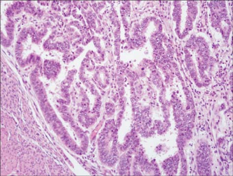
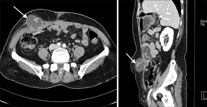
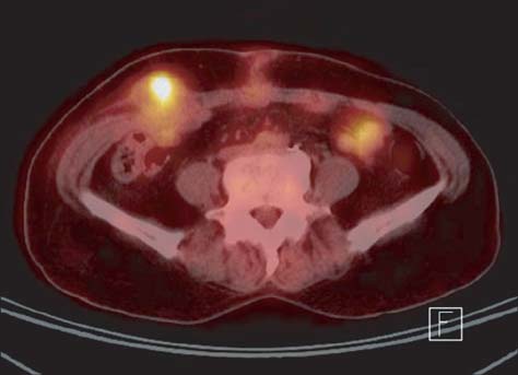
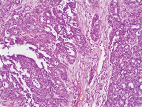
- Uterine papillary serous carcinoma (UPSC) is an aggressive form of endometrial cancer characterized by a high recurrence rate and poor prognosis. We report a case of a 58-year-old post-menopausal woman with an abdominal wall metastasis in stage IA UPSC. After surgical staging, she did not receive additional adjuvant therapy. An egg sized palpable mass developed in the right lower abdomen after 8 months. Both Abdominopelvic computed tomography (CT) and positron emission tomography (PET)-CT revealed a metastatic lesion in the abdominal wall. Hence, surgical excision was performed. The pathological findings showed metastatic UPSC with clear resection margin. After the diagnosis of UPSC metastasis in the abdominal wall, she received chemotherapy utilizing paclitaxel and carboplatin. After 3 years, no evidence of recurrence was found. Therefore, we suggest that even when UPSC is confined to the endometrium without lymph node metastasis and without lymphovascular invasion, chemotherapy should be considered as a postoperative adjuvant therapy.
MeSH Terms
Figure
Reference
-
1. Goff BA, Kato D, Schmidt RA, Ek M, Ferry JA, Muntz HG, et al. Uterine papillary serous carcinoma: patterns of metastatic spread. Gynecol Oncol. 1994; 54:264–268.2. Carcangiu ML, Chambers JT. Uterine papillary serous carcinoma: a study on 108 cases with emphasis on the prognostic significance of associated endometrioid carcinoma, absence of invasion, and concomitant ovarian carcinoma. Gynecol Oncol. 1992; 47:298–305.3. Chan JK, Loizzi V, Youssef M, Osann K, Rutgers J, Vasilev SA, et al. Significance of comprehensive surgical staging in noninvasive papillary serous carcinoma of the endometrium. Gynecol Oncol. 2003; 90:181–185.4. Wilson TO, Podratz KC, Gaffey TA, Malkasian GD Jr, O'Brien PC, Naessens JM. Evaluation of unfavorable histologic subtypes in endometrial adenocarcinoma. Am J Obstet Gynecol. 1990; 162:418–423. discussion 23-6.5. Chambers JT, Merino M, Kohorn EI, Peschel RE, Schwartz PE. Uterine papillary serous carcinoma. Obstet Gynecol. 1987; 69:109–113.6. Wheeler DT, Bell KA, Kurman RJ, Sherman ME. Minimal uterine serous carcinoma: diagnosis and clinicopathologic correlation. Am J Surg Pathol. 2000; 24:797–806.7. Huh WK, Powell M, Leath CA 3rd, Straughn JM Jr, Cohn DE, Gold MA, et al. Uterine papillary serous carcinoma: comparisons of outcomes in surgical Stage I patients with and without adjuvant therapy. Gynecol Oncol. 2003; 91:470–475.8. Havrilesky LJ, Secord AA, Bae-Jump V, Ayeni T, Calingaert B, Clarke-Pearson DL, et al. Outcomes in surgical stage I uterine papillary serous carcinoma. Gynecol Oncol. 2007; 105:677–682.9. Lim P, Al Kushi A, Gilks B, Wong F, Aquino-Parsons C. Early stage uterine papillary serous carcinoma of the endometrium: effect of adjuvant whole abdominal radiotherapy and pathologic parameters on outcome. Cancer. 2001; 91:752–757.10. Sutton G, Axelrod JH, Bundy BN, Roy T, Homesley HD, Malfetano JH, et al. Whole abdominal radiotherapy in the adjuvant treatment of patients with stage III and IV endometrial cancer: a gynecologic oncology group study. Gynecol Oncol. 2005; 97:755–763.11. Zanotti KM, Belinson JL, Kennedy AW, Webster KD, Markman M. The use of paclitaxel and platinum-based chemotherapy in uterine papillary serous carcinoma. Gynecol Oncol. 1999; 74:272–277.12. Ramondetta L, Burke TW, Levenback C, Bevers M, Bodurka-Bevers D, Gershenson DM. Treatment of uterine papillary serous carcinoma with paclitaxel. Gynecol Oncol. 2001; 82:156–161.13. Gehrig PA. Uterine papillary serous carcinoma: a review. Expert Opin Pharmacother. 2007; 8:809–816.14. Kelly MG, O'Malley DM, Hui P, McAlpine J, Yu H, Rutherford TJ, et al. Improved survival in surgical stage I patients with uterine papillary serous carcinoma (UPSC) treated with adjuvant platinum-based chemotherapy. Gynecol Oncol. 2005; 98:353–359.
- Full Text Links
- Actions
-
Cited
- CITED
-
- Close
- Share
- Similar articles
-
- A Case of Postirradiation Uterine Papillary Serous Carcinoma
- A case of Primary Serous Papillary Carcinoma of the Peritoneum.
- A Case of Papillary Serous Carcinoma of the Peritoneum
- A case of minimal uterine serous carcinoma with distant lymph node metastasis without peritoneal dissemination
- Primary Serous Papillary Carcinoma of the Peritoneum: A Case Report


