Dual-Energy CT for Detection of Traumatic Bone Bruises in the Knee Joint
- Affiliations
-
- 1Department of Radiology, Wonkwang University Hospital, Iksan, Korea. juhngsk@wonkwang.ac.kr
- 2Institute of Wonkwang Medical Science, Iksan, Korea.
Abstract
- PURPOSE
To evaluate the diagnostic performance of dual-energy computed tomography (DECT) in detecting traumatic bone marrow lesions in patients with acute knee injury.
MATERIALS AND METHODS
Between August 2011 and June 2012, 22 patients presenting with an acute knee injury, including 4 patients who were referred for bilateral knee trauma, underwent DECT (80 kVp and 140 kVp) and MR imaging. DECT data were postprocessed using a three-dimensional, color-coded, virtual non-calcium technique (VNC). DECT data were graded by 2 blinded independent readers using a four-point system (1 = distinct bone marrow lesion, 2 = less distinct bone marrow lesion, 3 = equivocal, 4 = none) for 6 femoral and tibial regions and 2 patellar regions. Routine MR knee imaging served as the reference standard.
RESULTS
MR images showed bone bruises in 81 of 364 regions. The overall sensitivity, specificity, positive predictive value, and negative predictive value of DECT for bone bruises were 65.4%, 98.2%, 91.4%, and 90.8%, respectively, for Reader 1 and 70.3%, 93.6%, 76.0%, and 91.7%, respectively, for Reader 2. In particular, tibial bone bruises could be found more easily with better sensitivity (80.2%).
CONCLUSION
The color-coded VNC technique with reconstructions from the DECT maybe helpful in detecting traumatic bone bruises with moderate sensitivity and excellent specificity compared to MR imaging.
MeSH Terms
Figure
-
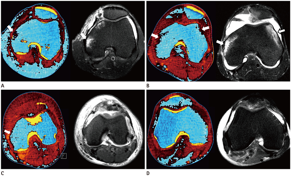
Fig. 1 Four-point grading system for axial color-coded VNC image (left) and corresponding T2-weighted STIR MR images (right). A. Grade 1: distinct bone marrow lesion, color-coded VNC images depict bone marrow abnormalities (arrow) in the same area as seen on the MR image (arrow). B. Grade 2: less distinct bone marrow lesion, "probable" bone marrow abnormality on color-coded VNC image (arrows). Corresponding MR image shows bone bruise in the same area (arrows). C. Grade 3: equivocal lesion, probably no bone marrow lesion on color-coded VNC image (arrow). MR image shows no bone bruise in medial femoral condyle. D. Grade 4: normal bone marrow, no abnormality on either modality.
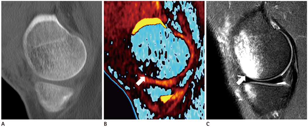
Fig. 2 A 23-year-old man with acute knee trauma. A. Sagittal weighted-average image simulating single-energy CT shows no fracture and normal trabecular structure. B. Color-coded VNC image shows bone bruise in the medial condyle of the femur (arrow). C. T2-weighted STIR MR image shows diffuse bone marrow lesion in the medial femoral condyle (arrow).
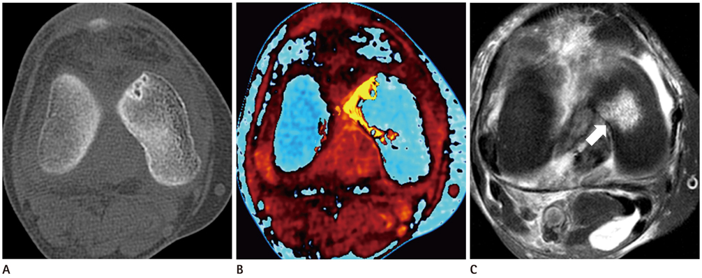
Fig. 3 A 72-year-old woman with acute knee trauma. A. Axial weighted-average image simulating single-energy CT shows no fracture. B. Color-coded VNC image shows no traumatic lesion in the medial femoral condyle. C. T2-weighted STIR MR image shows bone marrow edema in the medial femoral condyle (arrow).
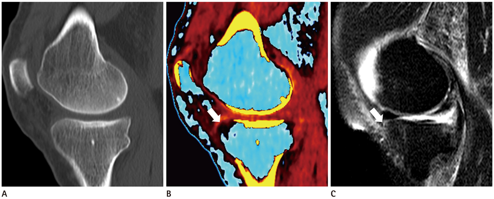
Fig. 4 A 61-year-old man with acute knee trauma. A. Sagittal weighted-average image simulating single-energy CT shows normal trabecular bone structure without fracture. B. Color-coded VNC image shows dark colored area in medial tibialplateau of knee joint (arrow). C. T2-weighted STIR MR image shows no bone bruise in medial tibial plateau (arrow).
Reference
-
1. Terzidis IP, Christodoulou AG, Ploumis AL, Metsovitis SR, Koimtzis M, Givissis P. The appearance of kissing contusion in the acutely injured knee in the athletes. Br J Sports Med. 2004; 38:592–596.2. Boks SS, Vroegindeweij D, Koes BW, Hunink MG, Bierma-Zeinstra SM. Follow-up of occult bone lesions detected at MR imaging: systematic review. Radiology. 2006; 238:853–862.3. Costa-Paz M, Muscolo DL, Ayerza M, Makino A, Aponte-Tinao L. Magnetic resonance imaging follow-up study of bone bruises associated with anterior cruciate ligament ruptures. Arthroscopy. 2001; 17:445–449.4. Sanders TG, Medynski MA, Feller JF, Lawhorn KW. Bone contusion patterns of the knee at MR imaging: footprint of the mechanism of injury. Radiographics. 2000; 20 Spec No:S135–S151.5. Vincken PW, Ter Braak BP, van Erkel AR, Coerkamp EG, Mallens WM, Bloem JL. Clinical consequences of bone bruise around the knee. Eur Radiol. 2006; 16:97–107.6. Nakamae A, Engebretsen L, Bahr R, Krosshaug T, Ochi M. Natural history of bone bruises after acute knee injury: clinical outcome and histopathological findings. Knee Surg Sports Traumatol Arthrosc. 2006; 14:1252–1258.7. Pache G, Krauss B, Strohm P, Saueressig U, Blanke P, Bulla S, et al. Dual-energy CT virtual noncalcium technique: detecting posttraumatic bone marrow lesions--feasibility study. Radiology. 2010; 256:617–624.8. Fischer MA, Gnannt R, Raptis D, Reiner CS, Clavien PA, Schmidt B, et al. Quantification of liver fat in the presence of iron and iodine: an ex-vivo dual-energy CT study. Invest Radiol. 2011; 46:351–358.9. Ryu KN, Jin W, Ko YT, Yoon Y, Oh JH, Park YK, et al. Bone bruises: MR characteristics and histological correlation in the young pig. Clin Imaging. 2000; 24:371–380.10. Zanetti M, Bruder E, Romero J, Hodler J. Bone marrow edema pattern in osteoarthritic knees: correlation between MR imaging and histologic findings. Radiology. 2000; 215:835–840.11. Geijer M, Dunker D, Collin D, Göthlin JH. Bone bruise, lipohemarthrosis, and joint effusion in CT of non-displaced hip fracture. Acta Radiol. 2012; 53:197–202.12. Guggenberger R, Gnannt R, Hodler J, Krauss B, Wanner GA, Csuka E, et al. Diagnostic performance of dual-energy CT for the detection of traumatic bone marrow lesions in the ankle: comparison with MR imaging. Radiology. 2012; 264:164–173.13. Mandalia V, Fogg AJ, Chari R, Murray J, Beale A, Henson JH. Bone bruising of the knee. Clin Radiol. 2005; 60:627–636.14. Mair SD, Schlegel TF, Gill TJ, Hawkins RJ, Steadman JR. Incidence and location of bone bruises after acute posterior cruciate ligament injury. Am J Sports Med. 2004; 32:1681–1687.15. Mandalia V, Henson JH. Traumatic bone bruising--a review article. Eur J Radiol. 2008; 67:54–61.16. Boks SS, Vroegindeweij D, Koes BW, Bernsen RM, Hunink MG, Bierma-Zeinstra SM. MRI follow-up of posttraumatic bone bruises of the knee in general practice. AJR Am J Roentgenol. 2007; 189:556–562.
- Full Text Links
-
- Actions
-
Cited
- CITED
-
- Close
- Share
-
- Similar articles
-
- Clinical Implications of Bone Bruises on MRI in Acute Traumatic ACL or PCL Injury
- An analysis of bone mineral contents using dual-energy computed tomography
- Dual-Energy CT: New Horizon in Medical Imaging
- Reconstuction of the Anterior and Posterior Cruciate Ligament Injury Associated with Traumatic Knee Joint Dislocation: Six Cases of Reconstruction Using Autogenous Achilles Tendon
- Dual-Layer Computed Tomography in Cardiovascular Imaging
