Dual-Energy CT: New Horizon in Medical Imaging
- Affiliations
-
- 1Department of Radiology and Research Institute of Radiology, Asan Medical Center, University of Ulsan College of Medicine, Seoul 05505, Korea. hwgoo@amc.seoul.kr
- 2Department of Radiology, Seoul National University College of Medicine, Seoul 03080, Korea.
- KMID: 2427225
- DOI: http://doi.org/10.3348/kjr.2017.18.4.555
Abstract
- Dual-energy CT has remained underutilized over the past decade probably due to a cumbersome workflow issue and current technical limitations. Clinical radiologists should be made aware of the potential clinical benefits of dual-energy CT over single-energy CT. To accomplish this aim, the basic principle, current acquisition methods with advantages and disadvantages, and various material-specific imaging methods as clinical applications of dual-energy CT should be addressed in detail. Current dual-energy CT acquisition methods include dual tubes with or without beam filtration, rapid voltage switching, dual-layer detector, split filter technique, and sequential scanning. Dual-energy material-specific imaging methods include virtual monoenergetic or monochromatic imaging, effective atomic number map, virtual non-contrast or unenhanced imaging, virtual non-calcium imaging, iodine map, inhaled xenon map, uric acid imaging, automatic bone removal, and lung vessels analysis. In this review, we focus on dual-energy CT imaging including related issues of radiation exposure to patients, scanning and post-processing options, and potential clinical benefits mainly to improve the understanding of clinical radiologists and thus, expand the clinical use of dual-energy CT; in addition, we briefly describe the current technical limitations of dual-energy CT and the current developments of photon-counting detector.
Keyword
MeSH Terms
Figure
-
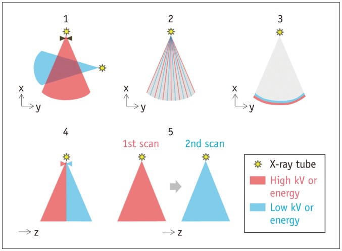
Fig. 1 Illustration of five different methods of dual-energy CT data acquisition.1 = dual tubes with or without beam filtration, 2 = rapid voltage switching with single tube, 3 = dual-layer detector with single tube, 4 = single tube with split filter, 5 = single tube with sequential dual scans
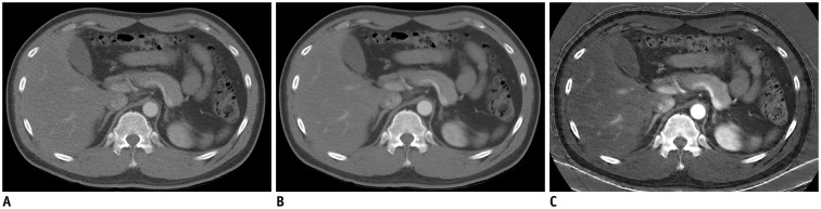
Fig. 2 Contrast-enhanced axial abdominal CT images using rapid voltage switching with single tube.A. Image generated immediately after dual-energy scanning by using 140 kVp projections only shows high image noise. B. Virtual monoenergetic image at 70 keV showing improved image quality needs to be additionally reconstructed for diagnostic imaging. C. Iodine map demonstrates improved iodine contrast-to-noise ratio. Of note, patient skin, cloth, and CT table appear artifactually bright on iodine map.
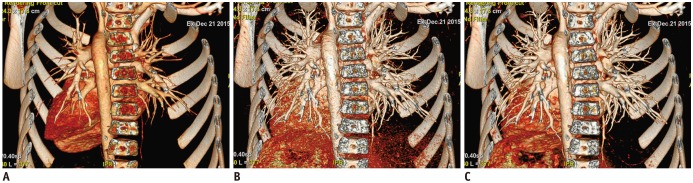
Fig. 3 Contrast-enhanced chest volume-rendered CT images with cropped posterior chest wall to unveil cardiovascular structures.A, B. Compared with volume-rendered image reconstructed from linearly mixed dual-energy images with ratio of 0.8 (A), volume-rendered 40 keV virtual monoenergetic image (B) shows further increase in cardiovascular opacification, but simultaneously increased noise compromises iodine contrast-to-noise ratio and image quality. C. On volume-rendered noise-optimized 40 keV virtual monoenergetic image, image noise reduction decoupled with increased iodine contrast leads to improved iodine contrast-to-noise ratio and image quality.
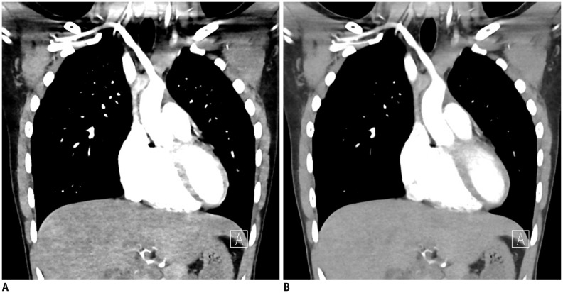
Fig. 4 Coronal chest noise-optimized virtual monoenergetic dual-energy CT imaging.Beam-hardening and/or photon starvation artifacts in thoracic inlet and shoulder pronounced in 40 keV image (A) are reduced in 60 keV images (B). Because iodine contrast is progressively reduced at higher keV images, overall optimal image quality can be achieved around 60 keV depending on patients' size as well as body region.
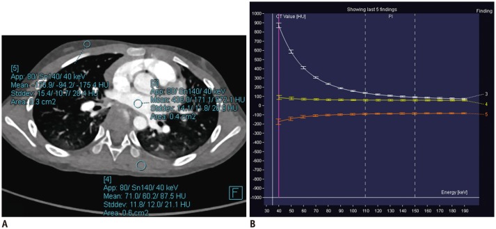
Fig. 5 Contrast-enhanced axial chest virtual monoenergetic dual-energy CT imaging.A. Three round regions of interest are placed in left atrium, back muscle, and subcutaneous fat in anterior chest wall, respectively, on axial chest CT image. B. Graph illustrating changes in CT value in three regions of interest as function of energy. Iodine in blood (white line) shows higher CT values at lower keV, while fat (orange line) reveals lower CT values at lower keV. In contrast, muscle (yellow line) demonstrates almost constant CT values in range of 40–190 keV.
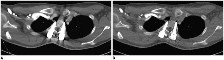
Fig. 6 Contrast-enhanced axial chest dual-energy CT imaging with posterior spinal fixation for scoliosis.A. Linearly mixed image with ratio of 0.4 shows beam-hardening artifacts caused by pedicle screws. B. Beam-hardening artifacts become less prominent on 130 keV image at expense of reduced iodine enhancement in vessels.
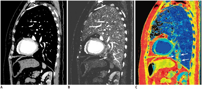
Fig. 7 Contrast-enhanced sagittal chest dual-energy CT imaging acquired with dual-layer detector technique.A. 70 keV image reveals subsegmental embolus (arrow) in anterior basal segment of left lower lobe. B, C. Wedge-shaped perfusion defect (arrows) is seen on iodine map (B) and more conspicuously on effective atomic number map (C).
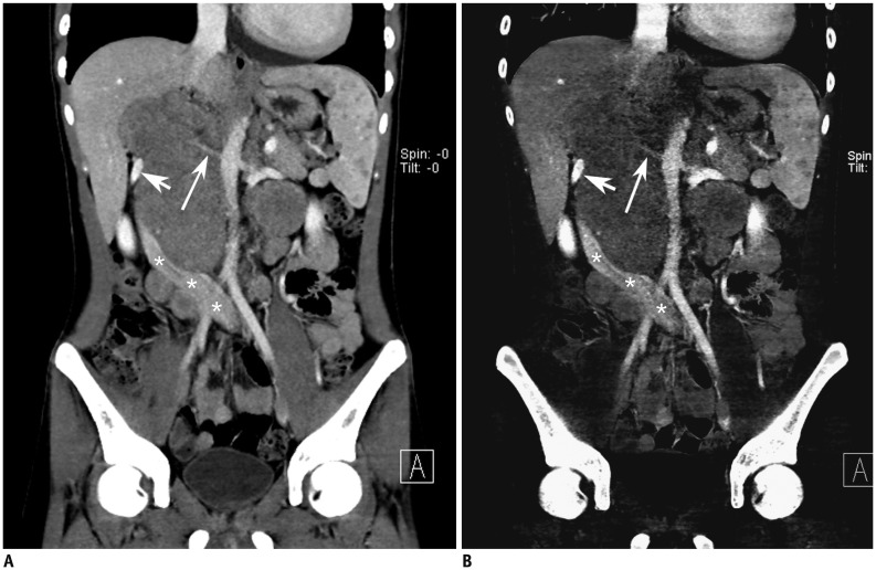
Fig. 8 Coronal abdominopelvic dual-energy CT imaging in patient with Hodgkin lymphoma.A, B. Linearly mixed image, iodine map. Right renal artery (long arrows), left renal vein (short arrows), inferior vena cava (asterisks) are displaced or encased by extensive, necrotic retroperitoneal lymphadenopathy. Lymphadenopathy shows subtle peripheral enhancement on iodine map (B). Multiple hypodense small nodules are noted in spleen.
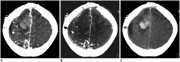
Fig. 9 Axial brain dual-energy CT imaging in patient with recurrent primitive neuroectodermal tumor.A-C. Linearly mixed image, iodine map, virtual non-contrast image. Larger anterior hyperdense lesion, pure intracerebral hemorrhage, shows no enhancement on iodine map (B) and hyperdensity suggesting recent hemorrhage on virtual non-contrast image (C). In contrast, smaller heterogenous lesion (arrows) reveals enhancing areas suggesting viable tumor on iodine map (B).
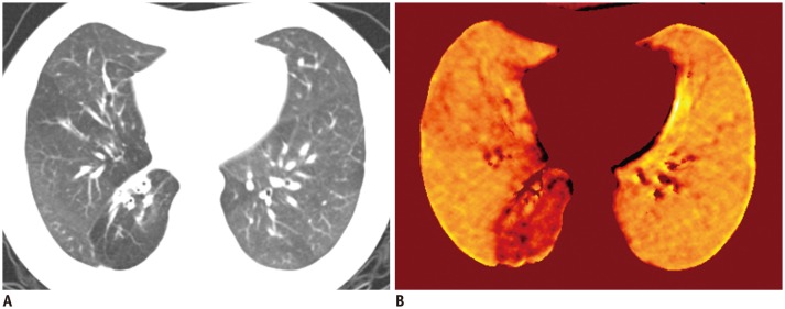
Fig. 10 Axial chest xenon-inhaled dual-energy CT imaging in patient with post-infectious bronchiolitis obliterans.A. Linearly mixed image shows bronchial wall thickenings and mosaic lung hyperlucency in right middle and lower lobes. Collapse of anterior basal segment of right lower lobe is also noted. B. Xenon map demonstrates severely reduced xenon enhancement in right lower lobe and mildly, heterogeneously decreased xenon enhancement in right middle lobe.
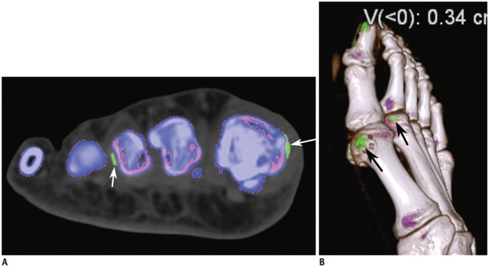
Fig. 11 Dual-energy CT imaging of right foot in patient with gout.Color-coded map (A) and volume-rendered image (B) show periarticular green foci (arrows) suggesting monosodium urate deposits and associated soft tissue swelling. False-positive artifacts are noted in typical location around nail bed and skin of great toe on volume-rendered image (B).
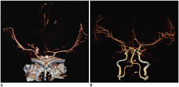
Fig. 12 Head CT angiographic volume-rendered imaging.A. Three-dimensional dual-energy angiographic image after automatic dual-energy bone removal shows residual bone at skull base due to incomplete dual-energy iodine-bone separation. B. Three-dimensional dual-energy angiographic image after detailed manual bone removal improves quality of head angiography but is time-consuming.
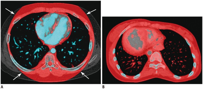
Fig. 13 Dual-energy chest CT imaging demonstrating lung vessels analysis.A. Axial image with lung vessels analysis shows normal enhancing pulmonary vessels in light blue in both lungs and limited dual-energy field of view (arrows) typically seen in dual-energy technique using dual X-ray tubes. B. In patient with dextrocaria, pulmonary atresia, ventricular septal defect, right aortic arch, and Eisenmenger syndrome, unobstructed pulmonary vessels in both lungs are red, secondary to very slow pulmonary circulation.
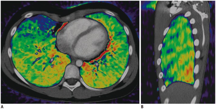
Fig. 14 Dual-energy pulmonary blood volume map demonstrating cardiac motion and beam-hardening artifacts.A. On axial pulmonary blood volume map, cardiac motion artifacts appear as red color areas around heart as well as blue areas in right middle lobe. B. On sagittal pulmonary blood volume map, beam-hardening artifacts appear as pattern of oblique stripes parallel to ribs.
Cited by 6 articles
-
Two Small Intravenous Catheters for High-Rate Contrast Medium Injection for Computed Tomography in Patients Lacking Superficial Veins to Accommodate a Large Catheter
Bum Gu Son, Min Jung Kim, Myeung Hwa Park, Kyoungsook Kim, Jiyu Kim, Se-Young Kim, Kyung Jin Lee, Sang Hyun Choi, Ah Young Kim, Seong Ho Park
Korean J Radiol. 2018;19(3):489-497. doi: 10.3348/kjr.2018.19.3.489.Iodine Quantification on Spectral Detector-Based Dual-Energy CT Enterography: Correlation with Crohn's Disease Activity Index and External Validation
Yeon Soo Kim, Se Hyung Kim, Hwa Sung Ryu, Joon Koo Han
Korean J Radiol. 2018;19(6):1077-1088. doi: 10.3348/kjr.2018.19.6.1077.Optimal Monochromatic Imaging of Spectral Computed Tomography Potentially Improves the Quality of Hepatic Vascular Imaging
Xiao-Ping Yin, Bu-Lang Gao, Cai-Ying Li, Huan Zhou, Liang Zhao, Ya-Ting Zheng, Yong-Xia Zhao
Korean J Radiol. 2018;19(4):578-584. doi: 10.3348/kjr.2018.19.4.578.Comparison of Filtered Back Projection, Hybrid Iterative Reconstruction, Model-Based Iterative Reconstruction, and Virtual Monoenergetic Reconstruction Images at Both Low- and Standard-Dose Settings in Measurement of Emphysema Volume and Airway Wall Thickness: A CT Phantom Study
Cherry Kim, Ki Yeol Lee, Chol Shin, Eun-Young Kang, Yu-Whan Oh, Moin Ha, Chang Sub Ko, Jaehyung Cha
Korean J Radiol. 2018;19(4):809-817. doi: 10.3348/kjr.2018.19.4.809.User-Friendly Vendor-Specific Guideline for Pediatric Cardiothoracic Computed Tomography Provided by the Asian Society of Cardiovascular Imaging Congenital Heart Disease Study Group: Part 1. Imaging Techniques
Sun Hwa Hong, Hyun Woo Goo, Eriko Maeda, Ki Seok Choo, I-Chen Tsai,
Korean J Radiol. 2019;20(2):190-204. doi: 10.3348/kjr.2018.0571.Optimal Kiloelectron Volt for Noise-Optimized Virtual Monoenergetic Images of Dual-Energy Pediatric Abdominopelvic Computed Tomography: Preliminary Results
Taek Min Kim, Young Hun Choi, Jung-Eun Cheon, Woo Sun Kim, In-One Kim, Ji Eun Park, Su-mi Shin, Seong Yong Pak, Bernhard Krauss
Korean J Radiol. 2019;20(2):283-294. doi: 10.3348/kjr.2017.0507.
Reference
-
1. Goo HW. CT radiation dose optimization and estimation: an update for radiologists. Korean J Radiol. 2012; 13:1–11. PMID: 22247630.
Article2. Johnson TR, Krauss B, Sedlmair M, Grasruck M, Bruder H, Morhard D, et al. Material differentiation by dual energy CT: initial experience. Eur Radiol. 2007; 17:1510–1517. PMID: 17151859.
Article3. Hounsfield GN. Computerized transverse axial scanning (tomography). 1. Description of system. Br J Radiol. 1973; 46:1016–1022. PMID: 4757352.4. Johnson TR. Dual-energy CT: general principles. AJR Am J Roentgenol. 2012; 199(5 Suppl):S3–S8. PMID: 23097165.
Article5. McCollough CH, Leng S, Yu L, Fletcher JG. Dual- and multi-energy CT: principles, technical approaches, and clinical applications. Radiology. 2015; 276:637–653. PMID: 26302388.
Article6. Maturen KE, Kaza RK, Liu PS, Quint LE, Khalatbari SH, Platt JF. “Sweet spot” for endoleak detection: optimizing contrast to noise using low keV reconstructions from fast-switch kVp dual-energy CT. J Comput Assist Tomogr. 2012; 36:83–87. PMID: 22261775.7. Faby S, Kuchenbecker S, Sawall S, Simons D, Schlemmer HP, Lell M, et al. Performance of today's dual energy CT and future multi energy CT in virtual non-contrast imaging and in iodine quantification: a simulation study. Med Phys. 2015; 42:4349–4366. PMID: 26133632.
Article8. Mileto A, Barina A, Marin D, Stinnett SS, Roy Choudhury K, Wilson JM, et al. Virtual monochromatic images from dual-energy multidetector CT: variance in CT numbers from the same lesion between single-source projection-based and dual-source image-based implementations. Radiology. 2016; 279:269–277. PMID: 26536403.
Article9. Yu L, Leng S, McCollough CH. Dual-energy CT-based monochromatic imaging. AJR Am J Roentgenol. 2012; 199(5 Suppl):S9–S15. PMID: 23097173.
Article10. Leng S, Yu L, Fletcher JG, McCollough CH. Maximizing iodine contrast-to-noise ratios in abdominal CT imaging through use of energy domain noise reduction and virtual monoenergetic dual-energy CT. Radiology. 2015; 276:562–570. PMID: 25860839.
Article11. Albrecht MH, Trommer J, Wichmann JL, Scholtz JE, Martin SS, Lehnert T, et al. Comprehensive comparison of virtual monoenergetic and linearly blended reconstruction techniques in third-generation dual-source dual-energy computed tomography angiography of the thorax and abdomen. Invest Radiol. 2016; 51:582–590. PMID: 26953565.
Article12. Wichmann JL, Gillott MR, De Cecco CN, Mangold S, Varga-Szemes A, Yamada R, et al. Dual-energy computed tomography angiography of the lower extremity runoff: impact of noise-optimized virtual monochromatic imaging on image quality and diagnostic accuracy. Invest Radiol. 2016; 51:139–146. PMID: 26561048.13. Pomerantz SR, Kamalian S, Zhang D, Gupta R, Rapalino O, Sahani DV, et al. Virtual monochromatic reconstruction of dual-energy unenhanced head CT at 65-75 keV maximizes image quality compared with conventional polychromatic CT. Radiology. 2013; 266:318–325. PMID: 23074259.
Article14. Agrawal MD, Pinho DF, Kulkarni NM, Hahn PF, Guimaraes AR, Sahani DV. Oncologic applications of dual-energy CT in the abdomen. Radiographics. 2014; 34:589–612. PMID: 24819783.
Article15. Bongers MN, Schabel C, Thomas C, Raupach R, Notohamiprodjo M, Nikolaou K, et al. Comparison and combination of dual-energy- and iterative-based metal artefact reduction on hip prosthesis and dental implants. PLoS One. 2015; 10:e0143584. PMID: 26600188.
Article16. Garcia LI, Azorin JF, Almansa JF. A new method to measure electron density and effective atomic number using dual-energy CT images. Phys Med Biol. 2016; 61:265–279. PMID: 26649484.
Article17. Chen CY, Hsu JS, Jaw TS, Shih MC, Lee LJ, Tsai TH, et al. Split-bolus portal venous phase dual-energy CT urography: protocol design, image quality, and dose reduction. AJR Am J Roentgenol. 2015; 205:W492–W501. PMID: 26496571.
Article18. De Cecco CN, Darnell A, Rengo M, Muscogiuri G, Bellini D, Ayuso C, et al. Dual-energy CT: oncologic applications. AJR Am J Roentgenol. 2012; 199(5 Suppl):S98–S105. PMID: 23097174.
Article19. Chae EJ, Song JW, Seo JB, Krauss B, Jang YM, Song KS. Clinical utility of dual-energy CT in the evaluation of solitary pulmonary nodules: initial experience. Radiology. 2008; 249:671–681. PMID: 18796658.
Article20. Lee HA, Lee YH, Yoon KH, Bang DH, Park DE. Comparison of virtual unenhanced images derived from dual-energy CT with true unenhanced images in evaluation of gallstone disease. AJR Am J Roentgenol. 2016; 206:74–80. PMID: 26700337.
Article21. Krauss B, Grant KL, Schmidt BT, Flohr TG. The importance of spectral separation: an assessment of dual-energy spectral separation for quantitative ability and dose efficiency. Invest Radiol. 2015; 50:114–118. PMID: 25373305.22. Luo XF, Xie XQ, Cheng S, Yang Y, Yan J, Zhang H, et al. Dual-energy CT for patients suspected of having liver iron overload: can virtual iron content imaging accurately quantify liver iron content? Radiology. 2015; 277:95–103. PMID: 25880263.
Article23. Omoumi P, Verdun FR, Guggenberger R, Andreisek G, Becce F. Dual-energy CT: basic principles, technical approaches, and applications in musculoskeletal imaging (part 2). Semin Musculoskelet Radiol. 2015; 19:438–445. PMID: 26696082.
Article24. Pache G, Krauss B, Strohm P, Saueressig U, Blanke P, Bulla S, et al. Dual-energy CT virtual noncalcium technique: detecting posttraumatic bone marrow lesions--feasibility study. Radiology. 2010; 256:617–624. PMID: 20551186.
Article25. McLaughlin PD, Mallinson P, Lourenco P, Nicolaou S. Dual-energy computed tomography: advantages in the acute setting. Radiol Clin North Am. 2015; 53:619–638. viiPMID: 26046502.26. Thieme SF, Johnson TR, Lee C, McWilliams J, Becker CR, Reiser MF, et al. Dual-energy CT for the assessment of contrast material distribution in the pulmonary parenchyma. AJR Am J Roentgenol. 2009; 193:144–149. PMID: 19542406.
Article27. Goo HW. Initial experience of dual-energy lung perfusion CT using a dual-source CT system in children. Pediatr Radiol. 2010; 40:1536–1544. PMID: 20596701.
Article28. Otrakji A, Digumarthy SR, Lo Gullo R, Flores EJ, Shepard JA, Kalra MK. Dual-energy CT: spectrum of thoracic abnormalities. Radiographics. 2016; 36:38–52. PMID: 26761530.
Article29. Hong YJ, Kim JY, Choe KO, Hur J, Lee HJ, Choi BW, et al. Different perfusion pattern between acute and chronic pulmonary thromboembolism: evaluation with two-phase dual-energy perfusion CT. AJR Am J Roentgenol. 2013; 200:812–817. PMID: 23521453.
Article30. Iyer KS, Newell JD Jr, Jin D, Fuld MK, Saha PK, Hansdottir S, et al. Quantitative dual-energy computed tomography supports a vascular etiology of smoking-induced inflammatory lung disease. Am J Respir Crit Care Med. 2016; 193:652–661. PMID: 26569033.
Article31. Baxa J, Matouskova T, Krakorova G, Schmidt B, Flohr T, Sedlmair M, et al. Dual-phase dual-energy CT in patients treated with erlotinib for advanced non-small cell lung cancer: possible benefits of iodine quantification in response assessment. Eur Radiol. 2016; 26:2828–2836. PMID: 26563350.
Article32. Kim SJ, Lim HK, Lee HY, Choi CG, Lee DH, Suh DC, et al. Dual-energy CT in the evaluation of intracerebral hemorrhage of unknown origin: differentiation between tumor bleeding and pure hemorrhage. AJNR Am J Neuroradiol. 2012; 33:865–872. PMID: 22241388.
Article33. Tijssen MP, Hofman PA, Stadler AA, van Zwam W, de Graaf R, van Oostenbrugge RJ, et al. The role of dual energy CT in differentiating between brain haemorrhage and contrast medium after mechanical revascularisation in acute ischaemic stroke. Eur Radiol. 2014; 24:834–840. PMID: 24258277.
Article34. Jin KN, De Cecco CN, Caruso D, Tesche C, Spandorfer A, Varga-Szemes A, et al. Myocardial perfusion imaging with dual energy CT. Eur J Radiol. 2016; 85:1914–1921. PMID: 27427412.
Article35. Hur J, Kim YJ, Lee HJ, Nam JE, Hong YJ, Kim HY, et al. Cardioembolic stroke: dual-energy cardiac CT for differentiation of left atrial appendage thrombus and circulatory stasis. Radiology. 2012; 263:688–695. PMID: 22495682.
Article36. Ascenti G, Mazziotti S, Lamberto S, Bottari A, Caloggero S, Racchiusa S, et al. Dual-energy CT for detection of endoleaks after endovascular abdominal aneurysm repair: usefulness of colored iodine overlay. AJR Am J Roentgenol. 2011; 196:1408–1414. PMID: 21606306.
Article37. Goo HW, Chae EJ, Seo JB, Hong SJ. Xenon ventilation CT using a dual-source dual-energy technique: dynamic ventilation abnormality in a child with bronchial atresia. Pediatr Radiol. 2008; 38:1113–1116. PMID: 18542942.
Article38. Chae EJ, Seo JB, Goo HW, Kim N, Song KS, Lee SD, et al. Xenon ventilation CT with a dual-energy technique of dual-source CT: initial experience. Radiology. 2008; 248:615–624. PMID: 18641254.
Article39. Park EA, Goo JM, Park SJ, Lee HJ, Lee CH, Park CM, et al. Chronic obstructive pulmonary disease: quantitative and visual ventilation pattern analysis at xenon ventilation CT performed by using a dual-energy technique. Radiology. 2010; 256:985–997. PMID: 20651060.
Article40. Chae EJ, Seo JB, Lee J, Kim N, Goo HW, Lee HJ, et al. Xenon ventilation imaging using dual-energy computed tomography in asthmatics: initial experience. Invest Radiol. 2010; 45:354–361. PMID: 20404734.41. Goo HW, Yu J. Redistributed regional ventilation after the administration of a bronchodilator demonstrated on xenon-inhaled dual-energy CT in a patient with asthma. Korean J Radiol. 2011; 12:386–389. PMID: 21603299.
Article42. Kim WW, Lee CH, Goo JM, Park SJ, Kim JH, Park EA, et al. Xenon-enhanced dual-energy CT of patients with asthma: dynamic ventilation changes after methacholine and salbutamol inhalation. AJR Am J Roentgenol. 2012; 199:975–981. PMID: 23096168.
Article43. Goo HW, Yang DH, Hong SJ, Yu J, Kim BJ, Seo JB, et al. Xenon ventilation CT using dual-source and dual-energy technique in children with bronchiolitis obliterans: correlation of xenon and CT density values with pulmonary function test results. Pediatr Radiol. 2010; 40:1490–1497. PMID: 20411254.
Article44. Goo HW, Yang DH, Kim N, Park SI, Kim DK, Kim EA. Collateral ventilation to congenital hyperlucent lung lesions assessed on xenon-enhanced dynamic dual-energy CT: an initial experience. Korean J Radiol. 2011; 12:25–33. PMID: 21228937.
Article45. Honda N, Osada H, Watanabe W, Nakayama M, Nishimura K, Krauss B, et al. Imaging of ventilation with dual-energy CT during breath hold after single vital-capacity inspiration of stable xenon. Radiology. 2012; 262:262–268. PMID: 22025733.
Article46. Goo HW. Dual-energy lung perfusion and ventilation CT in children. Pediatr Radiol. 2013; 43:298–307. PMID: 23417255.
Article47. Yoon SH, Goo JM, Jung J, Hong H, Park EA, Lee CH, et al. Computer-aided classification of visual ventilation patterns in patients with chronic obstructive pulmonary disease at two-phase xenon-enhanced CT. Korean J Radiol. 2014; 15:386–396. PMID: 24843245.
Article48. Hachulla AL, Pontana F, Wemeau-Stervinou L, Khung S, Faivre JB, Wallaert B, et al. Krypton ventilation imaging using dual-energy CT in chronic obstructive pulmonary disease patients: initial experience. Radiology. 2012; 263:253–259. PMID: 22332068.
Article49. Hong SR, Chang S, Im DJ, Suh YJ, Hong YJ, Hur J, et al. Feasibility of single scan for simultaneous evaluation of regional krypton and iodine concentrations with dual-energy CT: an experimental study. Radiology. 2016; 281:597–605. PMID: 27203543.50. Qu M, Ramirez-Giraldo JC, Leng S, Williams JC, Vrtiska TJ, Lieske JC, et al. Dual-energy dual-source CT with additional spectral filtration can improve the differentiation of non-uric acid renal stones: an ex vivo phantom study. AJR Am J Roentgenol. 2011; 196:1279–1287. PMID: 21606290.
Article51. Li X, Zhao R, Liu B, Yu Y. Gemstone spectral imaging dual-energy computed tomography: a novel technique to determine urinary stone composition. Urology. 2013; 81:727–730. PMID: 23453078.
Article52. Coupal TM, Mallinson PI, Gershony SL, McLaughlin PD, Munk PL, Nicolaou S, et al. Getting the most from your dual-energy scanner: recognizing, reducing, and eliminating artifacts. AJR Am J Roentgenol. 2016; 206:119–128. PMID: 26700343.
Article53. Schulz B, Kuehling K, Kromen W, Siebenhandl P, Kerl MJ, Vogl TJ, et al. Automatic bone removal technique in whole-body dual-energy CT angiography: performance and image quality. AJR Am J Roentgenol. 2012; 199:W646–W650. PMID: 23096210.
Article54. Lee CW, Seo JB, Song JW, Kim MY, Lee HY, Park YS, et al. Evaluation of computer-aided detection and dual energy software in detection of peripheral pulmonary embolism on dual-energy pulmonary CT angiography. Eur Radiol. 2011; 21:54–62. PMID: 20680290.
Article55. Atak H, Shikhaliev PM. Dual energy CT with photon counting and dual source systems: comparative evaluation. Phys Med Biol. 2015; 60:8949–8975. PMID: 26539971.
Article
- Full Text Links
-
- Actions
-
Cited
- CITED
-
- Close
- Share
-
- Similar articles
-
- Dual-Layer Computed Tomography in Cardiovascular Imaging
- Spectral Computed Tomography: Fundamental Principles and Recent Developments
- Analysis of Beam Hardening of Modulation Layers for Dual Energy Cone-beam CT
- Redistributed Regional Ventilation after the Administration of a Bronchodilator Demonstrated on Xenon-Inhaled Dual-Energy CT in a Patient with Asthma
- Clinical Applications of Dual-Energy CT
