A Rare Case of Bronchial Epithelial-Myoepithelial Carcinoma with Solid Lobular Growth in a 53-Year-Old Woman
- Affiliations
-
- 1Department of Pathology, Samsung Medical Center, Sungkyunkwan University School of Medicine, Seoul, Korea. hanjho@skku.edu
- 2Department of Radiology, Samsung Medical Center, Sungkyunkwan University School of Medicine, Seoul, Korea.
- 3Division of Pulmonary and Critical Care Medicine, Samsung Medical Center, Sungkyunkwan University School of Medicine, Seoul, Korea.
- 4Department of Thoracic Surgery, Samsung Medical Center, Sungkyunkwan University School of Medicine, Seoul, Korea.
- KMID: 2320721
- DOI: http://doi.org/10.4046/trd.2015.78.4.428
Abstract
- Epithelial-myoepithelial carcinoma (EMC) of lung is a minor subset of salivary type carcinoma of lung of known low grade malignancy. Histologically, two-cell components forming duct-like structure with inner epithelial cell layer and outer myoepithelial cell layer are characteristics of EMC. In salivary gland, dedifferentiation of conventional low grade malignancy has been reported and is thought to be related with poor prognosis. However, precise histomorphology and prognostic factors of pulmonary EMC have not been clarified due to its rarity. Herein, we reported a rare case of EMC presented as endobronchial mass in a 53-year old woman, which showed predominant solid lobular growth pattern and lymph node metastases.
Keyword
MeSH Terms
Figure
-

Figure 1 (A) Chest computed tomography reveals endobronchial mass in right bronchus intermedius (arrowheads). (B) Bronchoscopy shows lobulated endobronchial mass. (C) Multifocal fluorodeoxyglucose uptake in right bronchus intermedius (arrow) and right paratracheal area (arrowhead) on preoperative positron emission tomography-computed tomography.
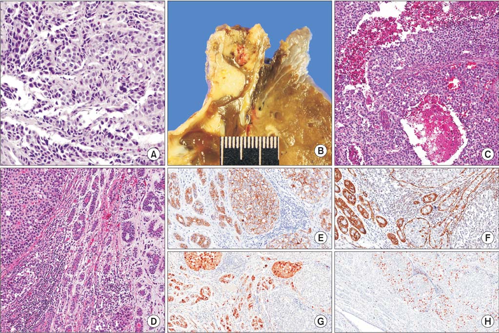
Figure 2 (A) Infiltrating atypical nests are identified on histologic examination of biopsy specimen (H&E stain, ×200). (B) Grossly, endobronchial yellow-tan solid mass focally interrupts the bronchial cartilage. (C) Tumor cells of solid lobular area demonstrate moderate cytologic atypia and discohesive pattern with accompanied multifocal central necrosis (H&E stain, ×100). (D) Toward the periphery of mass transition from duct-like two-cell layered area to the solid lobules is present (H&E stain, ×100). (E) Cytokeratin (CK) (AE1/AE3) is strong positive in tumor cells of inner layer of duct-like area. Outer layer of duct-like structure and solid area show variable intensity of CK (AE1/AE3) positivity (CK [AE1/AE3], ×100). (F) Smooth muscle actin (SMA) highlights the outer myoepithelial layer of duct-like area, which is only focally expressed in solid lobular area (SMA, ×100). (G) S-100 protein is positive in the outer myoepithelial layer of duct-forming area and variably expressed in solid lobular area (S-100 protein, ×100). (H) Ki-67 proliferative index is notably higher in periphery of solid lobular area, as compared to the center of solid lobules and adjacent duct-like structure (Ki-67, ×100).
Reference
-
1. Song DH, Choi IH, Ha SY, Han KM, Han J, Kim TS, et al. Epithelial-myoepthelial carcinoma of the tracheobronchial tree: the prognostic role of myoepithelial cells. Lung Cancer. 2014; 83:416–419.2. Zhu F, Liu Z, Hou Y, He D, Ge X, Bai C, et al. Primary salivary gland-type lung cancer: clinicopathological analysis of 88 cases from China. J Thorac Oncol. 2013; 8:1578–1584.3. Muslimani AA, Kundranda M, Jain S, Daw HA. Recurrent bronchial epithelial-myoepithelial carcinoma after local therapy. Clin Lung Cancer. 2007; 8:386–388.4. Nguyen CV, Suster S, Moran CA. Pulmonary epithelial-myoepithelial carcinoma: a clinicopathologic and immunohistochemical study of 5 cases. Hum Pathol. 2009; 40:366–373.5. Nishihara M, Takeda N, Tatsumi S, Kidoguchi K, Hayashi S, Sasayama T, et al. Skull metastasis as initial manifestation of pulmonary epithelial-myoepithelial carcinoma: a case report of an unusual case. Case Rep Oncol Med. 2011; 2011:610383.6. Gupta S, Bhalotra B, Jain N. Spectrum of intrabronchial mass lesions and role of flexible bronchoscopy in their diagnosis: a series of 74 cases. Indian J Chest Dis Allied Sci. 2010; 52:79–82.7. Pelosi G, Fraggetta F, Maffini F, Solli P, Cavallon A, Viale G. Pulmonary epithelial-myoepithelial tumor of unproven malignant potential: report of a case and review of the literature. Mod Pathol. 2001; 14:521–526.
- Full Text Links
-
- Actions
-
Cited
- CITED
-
- Close
- Share
-
- Similar articles
-
- Epithelial-myoepithelial carcinoma of the parotid gland: a case report
- Epithelial-myoepithelial carcinoma arising in pleomorphic adenoma of palate
- Malignant Adenomyoepithelioma of the Breast
- A Case of Epithelial-Myoepithelial Carcinoma in Left Submandibular Gland
- Epithelial-Myoepithelial Carcinoma of Intercalated Duct of Parotid Gland
