Prevalence of Benign Diseases Mimicking Lung Cancer: Experience from a University Hospital of Southern Brazil
- Affiliations
-
- 1Department of Pneumology, University Hospital of Santa Maria, Santa Maria, Brazil. guto.h@hotmail.com
- 2Porto Alegre Clinical Hospital, Porto Alegre, Brazil.
- 3Federal Univesity of Rio Grande do Sul, Porto Alegre, Brazil.
- 4Federal University of Santa Maria, Santa Maria, Brazil.
- KMID: 2320597
- DOI: http://doi.org/10.4046/trd.2015.78.2.72
Abstract
- BACKGROUND
Lung cancer is the most lethal type of cancer in the world. Several benign lung diseases may mimic lung carcinoma in its clinical and radiological presentation, which makes the differential diagnosis for granulomatous diseases more relevant in endemic regions like Brazil. This study was designed to describe the prevalence and the diagnostic work-up of benign diseases that mimic primary lung cancer in patients hospitalized at a university hospital from south of Brazil.
METHODS
This was a transversal study, which evaluated the medical records of 1,056 patients hospitalized for lung cancer treatment from September 2003 to September 2013 at University Hospital of Santa Maria.
RESULTS
Eight hundred and four patients underwent invasive procedures for suspected primary lung carcinoma. Primary lung cancer was confirmed in 77.4% of the patients. Benign disease was confirmed in 8% of all patients. Tuberculosis (n=14) and paracoccidioidomycosis (n=9) were the most frequent infectious diseases. The diagnosis of benign diseases was obtained by flexible bronchoscopy in 55.6% of the cases and by thoracotomy in 33.4%.
CONCLUSION
Infectious diseases are the most frequent benign diseases mimicking lung cancer at their initial presentation. Many of these cases could be diagnosed by minimally invasive procedures such as flexible bronchoscopy. Benign diseases should be included in the differential diagnosis during the investigation for primary lung cancer in order to avoid higher cost procedures and mortality.
MeSH Terms
Figure
-

Figure 1 Chest radiographs and chest and skull computed tomography (CT) scans of a 36-year-old male, smoker, with progressive dyspnea, weight loss, and productive cough. (A, B) Chest radiograph with opacity in the inferior lobe and in the left superior lobe. (C) Chest CT scan with voluminous lesion in the left lung. (D) Skull CT scan with nodular lesions in the brain hemispheres. Fibrobronchoscopy with biopsy of polypoid lesion in the bronchus of the left inferior lobe identified Cryptococcus sp. cultured with C. gatti growth.

Figure 2 Chest computed tomography (CT) scan of a 59-year-old patient, ex-smoker, retired farmer with evening fever for 2 weeks, weight loss, and pleuritic pain. Chest radiograph with nodular lesion in the left inferior lobe. Chest CT with juxta-pleural lesion in the left inferior lobe. A thoracotomy biopsy was performed. The histopathologic analysis showed granulomatous processes with areas of necrosis and fungus consistent with Paracoccidioides brasiliensis, confirmed by fungus growth in Sabouraud agar.
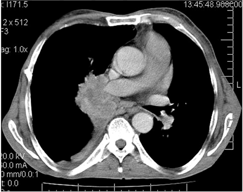
Figure 3 Chest computed tomography (CT) scan of a 70-year-old patient, retired farmer, smoker, alcoholic exhibits symptoms compatible with chronic obstructive pulmonary disease, progressing with loss of weight, cough, hemoptysis, and increased dyspnea. Chest radiograph with right perihilar opacity. Chest CT confirmed the finding. Fibrobronchoscopy with endobronchial biopsy of the lesion was performed and showed the presence of acid-fast bacilli, with confirmation of Mycobacterium tuberculosis in culture media.
Reference
-
1. Ferlay J, Soerjomataram I, Ervik M, Dikshit R, Eser S, Mathers C, et al. GLOBOCAN 2012 v1.0, cancer incidence and mortality worldwide: IARC CancerBase No. 11 [Internet]. Lyon: IARC Press;2013. cited 2013 Dec 20. Available from: http://globocan.iarc.fr.2. DATASUS. Indicators of morbidity: incidence of malignancies [Internet]. Brasilia: DATASUS;2011. cited 2013 Dec 20. Available from: http://tabnet.datasus.gov.br/cgi/idb2010/d05_08ufm.htm.3. Ost D, Fein AM, Feinsilver SH. Clinical practice: the solitary pulmonary nodule. N Engl J Med. 2003; 348:2535–2542.4. dos Santos JW, Andrade CF, Lopes TS, Londero AT. Pseudotumoral presentation of chronic pulmonary paracoccidioidomycosis: report of a case. Mycopathologia. 1996; 134:135–136.5. Silva GA, Brandao DF, Vianna EO, Sa Filho JB, Baddini-Martinez J. Cryptococcosis, silicosis, and tuberculous pseudotumor in the same pulmonary lobe. J Bras Pneumol. 2013; 39:620–626.6. Lopes AJ, Jansen U, Capone D, Neves DD, Jansen JM. Diagnosis of false pulmonary tumours. Pulmano RJ. 2005; 14:33–42.7. Smith MA, Battafarano RJ, Meyers BF, Zoole JB, Cooper JD, Patterson GA. Prevalence of benign disease in patients undergoing resection for suspected lung cancer. Ann Thorac Surg. 2006; 81:1824–1828.8. Rolston KV, Rodriguez S, Dholakia N, Whimbey E, Raad I. Pulmonary infections mimicking cancer: a retrospective, three-year review. Support Care Cancer. 1997; 5:90–93.9. Agarwal R, Srinivas R, Aggarwal AN. Parenchymal pseudotumoral tuberculosis: case series and systematic review of literature. Respir Med. 2008; 102:382–389.10. Machuca TN, Cardoso PF, Camargo SM, Signori L, Andrade CF, Moreira AL, et al. Surgical treatment of bronchial carcinoid tumors: a single-center experience. Lung Cancer. 2010; 70:158–162.11. Schweigert M, Dubecz A, Beron M, Ofner D, Stein HJ. Pulmonary infections imitating lung cancer: clinical presentation and therapeutical approach. Ir J Med Sci. 2013; 182:73–80.12. Dall Bello AG, Severo CB, Hochhegger B, Oliveira FM, Severo LC. Infection mimicking cancer : retrospective and prospective evaluation of mycosis and actinomycetous. Rev Patol Trop. 2013; 42:395–401.13. Portal of Health, Ministry of Health. Tuberculosis: epidemiological situation [Internet]. Brasilia: Ministry of Health;2014. cited 2014 Nov 23. Available from: http://portalsaude.saude.gov.br/.14. Bellissimo-Rodrigues F, Machado AA, Martinez R. Paracoccidioidomycosis epidemiological features of a 1,000-cases series from a hyperendemic area on the southeast of Brazil. Am J Trop Med Hyg. 2011; 85:546–550.15. Toomes H, Delphendahl A, Manke HG, Vogt-Moykopf I. The coin lesion of the lung: a review of 955 resected coin lesions. Cancer. 1983; 51:534–537.16. Dick R. Transthoracic image guided biopsy. Postgrad Med J. 1988; 64:544–551.17. Swanson SJ, Jaklitsch MT, Mentzer SJ, Bueno R, Lukanich JM, Sugarbaker DJ. Management of the solitary pulmonary nodule: role of thoracoscopy in diagnosis and therapy. Chest. 1999; 116:6 Suppl. 523S–524S.18. Allison RD, Vincent AL, Greene JN, Sandin RL, Field T. Infectious pulmonary nodules mimicking lung carcinoma. Infect Med. 2004; 21:181–186.19. Patel VK, Naik SK, Naidich DP, Travis WD, Weingarten JA, Lazzaro R, et al. A practical algorithmic approach to the diagnosis and management of solitary pulmonary nodules: part 1: radiologic characteristics and imaging modalities. Chest. 2013; 143:825–839.20. Wanke B, Aidê MA. Chapter 6. Paracoccidioidomicose. J Bras Pneumol. 2009; 35:1245–1249.
- Full Text Links
-
- Actions
-
Cited
- CITED
-
- Close
- Share
-
- Similar articles
-
- Diagnostic Values of Carcinoembryonic Antigen in Bronchial Washing Fluid
- Immunoglobulin G4-Related Lung Disease Mimicking Lung Cancer: Two Case Reports
- Usefulness and Comparison of 201Tl - chloride, 99mTc - MIBI, 99mTc(V) - DMSA Single Photon Emission Computed Tomography in Distinguishing Lung Cancer from Benign Lesion
- Resting Energy Expenditure in Patients with Lung Cancer
- First Evaluation of an Outbreak of Bovine Babesiosis and Anaplasmosis in Southern Brazil Using Multiplex PCR
