A Case of Metastatic Endobronchial Melanoma from an Unknown Primary Site
- Affiliations
-
- 1Department of Internal Medicine, Kyungpook National University School of Medicine, Daegu, Korea. kimch@knu.ac.kr
- 2Department of Nuclear Medicine, Kyungpook National University School of Medicine, Daegu, Korea.
Abstract
- Melanoma can occur as a metastasis within subcutaneous tissue, lymph nodes, or viscera without a detectable primary tumor. Among patients with metastatic melanoma of unknown primary lesion, those with endobronchial metastasis are exceedingly rare. Herein we report a case of an endobronchial and pulmonary metastasis in a patient with melanoma originating from an unknown primary site. The patient without a previous history of melanoma presented with blood-tinged sputum. Fiberoptic bronchoscopy revealed a black polypoid tumor obstructing the posterior basal segmental bronchus of the right lower lobe. A final diagnosis of the malignant melanoma was made based on an immunohistochemical study of the bronchoscopic biopsy specimen. Skin, ophthalmic, oral, and nasal examinations failed to identify occult primary lesions. Subsequent evaluation including positron emission tomography/computed tomography scans did not uncover any abnormalities other than the metastatic pulmonary melanoma. We also describe the characteristic bronchoscopic features of melanoma.
Keyword
MeSH Terms
Figure
-
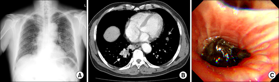
Figure 1 (A) Chest radiograph. Numerous small nodules were evenly scattered throughout both lungs with increased opacities toward the bilateral lower lobes. (B) Chest CT. Enhanced chest CT scan displayed a mass measuring 2×3 cm in the right lower lobe (white arrow) and another small nodule in the left lower lobe (gray arrow). (C) Bronchoscopic finding. A black polypoid tumor was found to be obstructing the posterior basal segmental bronchus of the right lower lobe. CT: computed tomography.
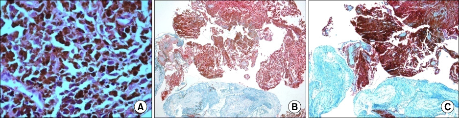
Figure 2 Histologic findings. Tumor cells with a prominent nucleus and brown pigment in the cytoplasm are shown (A, H&E stain, ×400). Immunohistochemical staining for S100 (B) and HMB45 (C) was positive.
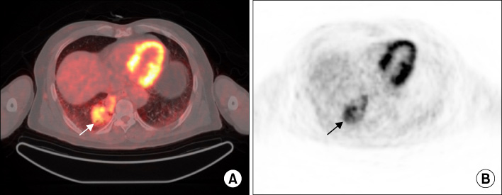
Figure 3 F-18 fluorodeoxyglucose PET/CT scan. PET/CT scan revealed a metabolically active mass (arrow) with a maximum standardized uptake value of 6.3 in the right lower lobe. (A) Transaxial fusion PET/CT image. (B) Transaxial PET image. PET: positron emission tomography; CT: computed tomography.
Reference
-
1. Dasgupta T, Bowden L, Berg JW. Malignant melanoma of unkonwn primary origin. Surg Gynecol Obstet. 1963. 117:341–345.2. Kamposioras K, Pentheroudakis G, Pectasides D, Pavlidis N. Malignant melanoma of unknown primary site. To make the long story short. A systematic review of the literature. Crit Rev Oncol Hematol. 2011. 78:112–126.3. Anbari KK, Schuchter LM, Bucky LP, Mick R, Synnestvedt M, Guerry D 4th, et al. University of Pennsylvania Pigmented Lesion Study Group. Melanoma of unknown primary site: presentation, treatment, and prognosis--a single institution study. Cancer. 1997. 79:1816–1821.4. Savoia P, Fava P, Osella-Abate S, Nardò T, Comessatti A, Quaglino P, et al. Melanoma of unknown primary site: a 33-year experience at the Turin Melanoma Centre. Melanoma Res. 2010. 20:227–232.5. Kelly J, Redmond HP. Melanoma of unknown origin: a case series. Ir J Med Sci. 2010. 179:629–632.6. Min YH, Kim SW, Chin HJ, Lee TY, Song HH, Lee KS, et al. Case of unknown primary malignant melanoma with pulmonary and endobronchial metastasis. Tuberc Respir Dis. 2002. 53:196–201.7. O'Neill JK, Khundar R, Knowles L, Scott-Young N, Orlando A. Melanoma with an unknown primary--a case series. J Plast Reconstr Aesthet Surg. 2010. 63:2071–2080.8. Gutzeit A, Antoch G, Kühl H, Egelhof T, Fischer M, Hauth E, et al. Unknown primary tumors: detection with dual-modality PET/CT--initial experience. Radiology. 2005. 234:227–234.9. Allen MS Jr, Drash EC. Primary melanoma of the lung. Cancer. 1968. 21:154–159.10. de Wilt JH, Farmer SE, Scolyer RA, McCaughan BC, Thompson JF. Isolated melanoma in the lung where there is no known primary site: metastatic disease or primary lung tumour? Melanoma Res. 2005. 15:531–537.11. Carstens PH, Kuhns JG, Ghazi C. Primary malignant melanomas of the lung and adrenal. Hum Pathol. 1984. 15:910–914.12. Shepherd MP. Endobronchial metastatic disease. Thorax. 1982. 37:362–365.13. Salud A, Porcel JM, Rovirosa A, Bellmunt J. Endobronchial metastatic disease: analysis of 32 cases. J Surg Oncol. 1996. 62:249–252.14. Koyi H, Brandén E. Intratracheal metastasis from malignant melanoma. J Eur Acad Dermatol Venereol. 2000. 14:407–408.15. Seam N, Khosla R. Metastatic endobronchial melanoma. Respiration. 2009. 77:214.
- Full Text Links
-
- Actions
-
Cited
- CITED
-
- Close
- Share
-
- Similar articles
-
- Malignant Melanoma of Unknown Primary Origin Presenting as Cardiac Metastasis
- A Case of Unknown Primary Malignant Melanoma with Pulmonary and Endobronchial Metastasis
- Unknow Primary Melanoma
- The Appearance of a Candidate Site for a Primary Melanoma: A 5 Year-gap with a Melanoma of an Unknown Site
- Two Cases of Malignant Melanoma with Nodal Metastasis from Unknown Primary Site
