First Isolation of Segniliparus rugosus from a Patient with Radiologic Features Similar to Non-Tuberculous Mycobacteriosis
- Affiliations
-
- 1Department of Internal Medicine, Seoul National University College of Medicine, Seoul, Korea.
- 2Department of Laboratory Medicine, National Cancer Center, Goyang, Korea.
- 3Center for Lung Cancer, National Cancer Center, Goyang, Korea. jekyde@yahoo.co.kr
Abstract
- In 2005, a group of mycolic acid-containing bacteria was characterized as belonging to a novel genus, Segniliparus with species Segniliparus rugosus and S. rotundus. We report a case of the S. rugosus isolated from a 54-year-old woman with radiologic features mimicking that of non-tuberculous mycobacteriosis (NTM). When the patient first visited our hospital, an acid-fast bacteria (AFB) smear tested positive and Mycobacterium tuberculosis polymerase chain reaction (TB PCR) was negative in the bronchoalveolar lavage sample. After 2 months, the growing colonies were reported as NTM, but could not be identified because they had died. One year after the initial visit, induced sputum samples showed the same results, positive AFB smear and negative TB PCR. At this point, the growing colonies were identified as S. rugosus. Therefore, we should consider Segniliparus genus as a differential diagnosis for AFB in respiratory specimens in addition to the genus Mycobacterium.
Keyword
MeSH Terms
Figure
-

Figure 1 Chest X-ray images. (A) Chest X-ray taken 7 months before the first OPD visit showed parenchymal infiltration in the left middle lung field. (B) Chest X-ray taken during the first visit showed persistent parenchymal infiltration in left middle lung field. OPD: outpatient department.
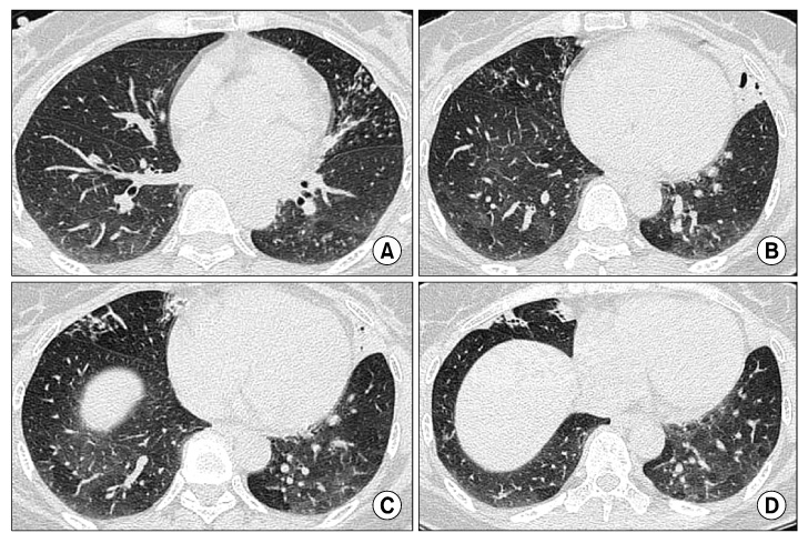
Figure 2 Chest HRCT images taken during the first visit revealed small nodules and branching centrilobular opacities in the left lingular segment (A), RML (B, C) and consolidation containing air bronchogram in the left lingular segment (B, C) and RML (D). HRCT: high resolution computed tomography; RML: right middle lobe.

Figure 3 Chest CT images taken 12 months after the first visit showed new branching centrilobular opacities in the left lower lobe (A, arrow) and persitent bronchiolitis and bronchiectatic features in the left lingular segment (B, C), RML (C, D). CT: computed tomography; RML: right middle lobe.

Figure 4 AFB stains (Ziehl-Neelsen stains) of induced sputum specimens (×1,000). AFB stains showed numerous acid fast bacilli, Segiliparus rugosus, which were later confirmed by 16s rRNA sequencing.
Reference
-
1. Butler WR, Floyd MM, Brown JM, Toney SR, Daneshvar MI, Cooksey RC, et al. Novel mycolic acid-containing bacteria in the family Segniliparaceae fam. nov., including the genus Segniliparus gen. nov., with descriptions of Segniliparus rotundus sp. nov. and Segniliparus rugosus sp. nov. Int J Syst Evol Microbiol. 2005. 55:1615–1624.2. Butler WR, Sheils CA, Brown-Elliott BA, Charles N, Colin AA, Gant MJ, et al. First isolations of Segniliparus rugosus from patients with cystic fibrosis. J Clin Microbiol. 2007. 45:3449–3452.3. Butler WR, Floyd MM, Silcox V, Cage G, Desmond E, Duffey PS, et al. Standardized method for HPLC identification of Mycobacteria. 1996. Atlanta, GA: CDC, US Department of Health and Human Services.4. Floyd MM, Gross WM, Bonato DA, Silcox VA, Smithwick RW, Metchock B, et al. Mycobacterium kubicae sp. nov., a slowly growing, scotochromogenic Mycobacterium. Int J Syst Evol Microbiol. 2000. 50:1811–1816.5. Griffith DE, Aksamit T, Brown-Elliott BA, Catanzaro A, Daley C, Gordin F, et al. An official ATS/IDSA statement: diagnosis, treatment, and prevention of nontuberculous mycobacterial diseases. Am J Respir Crit Care Med. 2007. 175:367–416.6. Hansen T, Van-Kerckhof J, Jelfs P, Wainwright C, Ryan P, Coulter C. Segniliparus rugosus infection, Australia. Emerg Infect Dis. 2009. 15:611–613.
- Full Text Links
-
- Actions
-
Cited
- CITED
-
- Close
- Share
-
- Similar articles
-
- A Case of Segniliparus rugosus Pulmonary Infection in an Immunocompetent Patient with Non-cystic Fibrosis
- Roentgenological evaluation of human atypical mycobacteriosis
- Mediastinal & pulmonary sarcoidosis : report of one case and radiologic-pathologic correlation of lymph node enhancement pattern during the treatment
- A Case of Primary Tuberculous Otitis Media in a 2-month Old Infant
- Delayed Diagnosis of Tuberculous Spondylitis Masked by Concomitant Methicillin Resistant Staphylococcus Aureus Infection
