A Case of Lymphoepithelioma-Like Carcinoma of the Lung
- Affiliations
-
- 1Department of Internal Medicine, Keimyung University School of Medicine, Daegu, Korea. jcy2475@dsmc.or.kr
- 2Department of Thoracic and Cardiovascular Surgery, and 3Pathology, Keimyung University School of Medicine, Daegu, Korea.
- 3Department of Pathology, Keimyung University School of Medicine, Daegu, Korea.
Abstract
- Lymphoepithelioma-like carcinoma (LELC) of the lung is a very rare tumor. Originally described in the nasopharynx as lymphoepithelioma, this carcinoma has also been found in the stomach, esophagus, thymus, cervix, urinary bladder, skin, and salivary glands. Histologically, it is an undifferentiated carcinoma that has a syncytial appearance with tumor cells and is infiltrated by numerous lymphocytes, macrophages, and plasma cells. LELC of the lung occurs more commonly in Asians, particularly Chinese. Many studies have reported the association between Epstein-Barr virus (EBV) and LELC of the lung in Asian patients. A 45-year-old man had a solitary pulmonary nodule on a routine chest X-ray examination. As a malignant tumor was suspected, surgical resection was performed to establish the correct diagnosis. The pathology of the excised tumor demonstrated LELC of the lung. This is the first report of LELC of the lung in Korea.
Keyword
MeSH Terms
Figure
-
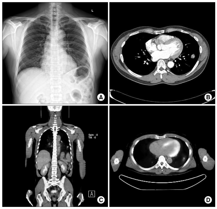
Figure 1 (A) Chest radiograph shows nodular lesion on the left lower lung field. (B) Chest CT scan shows a 2 cm sized nodule in the anterior basal segment of the left lower lobe. (C, D) F-18 FDG PET-CT scan shows hypermetabolic nodule in the left lower lobe (SUVmax, 6.1). CT: computed tomography; FDG: fluorodeoxyglucose; PET-CT: positron emission tomography-CT.
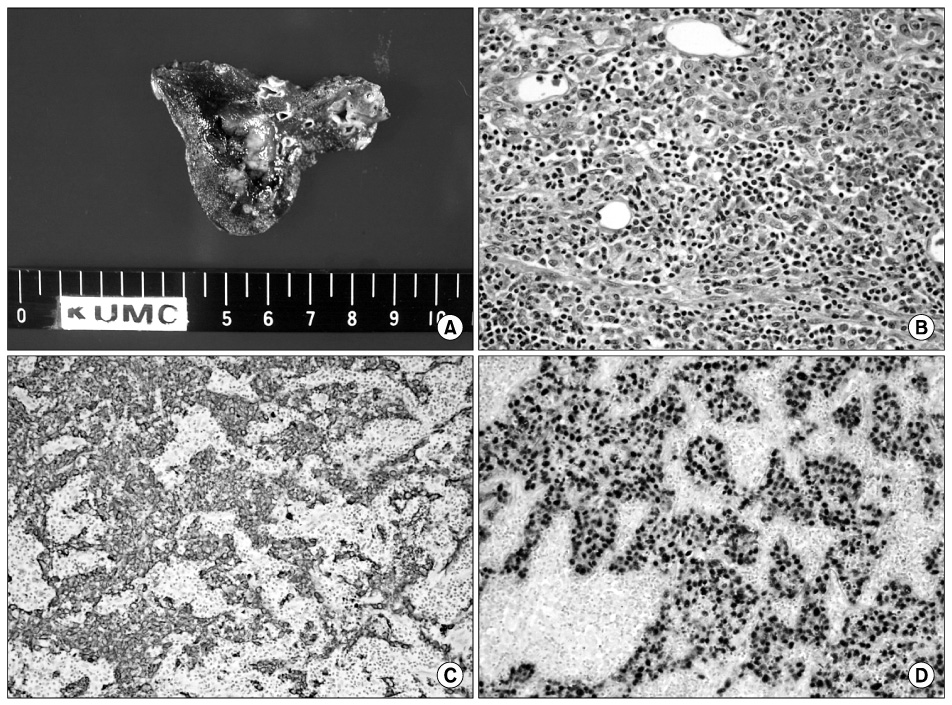
Figure 2 (A) Gross appearance reveals 2.5 cm sized round to oval nodule with ill demarcations in the left lower lobe. (B) H&E staining shows a sheet of tumor cells surrounded by abundant lymphoid cells in the stroma (×400). (C) Pan-CK stain shows stained tumor cells (epithelial origin), whereas lymphocytes are not (×200). (D) In situ hybridization for Epstein-Barr virus-encoded small nuclear RNA shows intense signals in tumor cell nuclei (dark blue) (EBV in situ, ×200).
Reference
-
1. Ho JC, Wong MP, Lam WK. Lymphoepithelioma-like carcinoma of the lung. Respirology. 2006. 11:539–545.2. Bégin LR, Eskandari J, Joncas J, Panasci L. Epstein-Barr virus related lymphoepithelioma-like carcinoma of lung. J Surg Oncol. 1987. 36:280–283.3. Han AJ, Xiong M, Zong YS. Association of Epstein-Barr virus with lymphoepithelioma-like carcinoma of the lung in southern China. Am J Clin Pathol. 2000. 114:220–226.4. Han AJ, Xiong M, Gu YY, Lin SX, Xiong M. Lymphoepithelioma-like carcinoma of the lung with a better prognosis. A clinicopathologic study of 32 cases. Am J Clin Pathol. 2001. 115:841–850.5. Chang YL, Wu CT, Shih JY, Lee YC. New aspects in clinicopathologic and oncogene studies of 23 pulmonary lymphoepithelioma-like carcinomas. Am J Surg Pathol. 2002. 26:715–723.6. Ho JC, Lam WK, Wong MP, Wong MK, Ooi GC, Ip MS, et al. Lymphoepithelioma-like carcinoma of the lung: experience with ten cases. Int J Tuberc Lung Dis. 2004. 8:890–895.7. Iezzoni JC, Gaffey MJ, Weiss LM. The role of Epstein-Barr virus in lymphoepithelioma-like carcinomas. Am J Clin Pathol. 1995. 103:308–315.8. Cho JH, Lee WS, Lee KR, Jeong HK, Cho SB, Joo YE, et al. Gastric lymphoepithelioma-like carcinoma diagnosed and treated by endoscopic submucosal dissection: review of the literature. Korean J Gastrointest Endosc. 2010. 40:256–260.9. Ki EY, Ro DY, Kim HJ, Park BJ, Kim YW, Kim TE, et al. A case of lymphoepithelioma-like carcinoma of the uterine cervix. Korean J Obstet Gynecol. 2009. 52:115–119.10. Yun HK, Yun SI, Lee YH, Kang KM, Kwak EK, Kim JS, et al. Lymphoepithelioma-like carcinoma of the urinary bladder. J Korean Med Sci. 2010. 25:1672–1675.11. Myung JH, Hong SK, Kim HH, Park BJ. A case of lymphoepithelioma like salivary gland carcinoma. J Clin Otolaryngol Head Neck Surg. 2006. 17:124–127.12. Ooi GC, Ho JC, Khong PL, Wong MP, Lam WK, Tsang KW. Computed tomography characteristics of advanced primary pulmonary lymphoepithelioma-like carcinoma. Eur Radiol. 2003. 13:522–526.13. Hoxworth JM, Hanks DK, Araoz PA, Elicker BM, Reddy GP, Webb WR, et al. Lymphoepithelioma-like carcinoma of the lung: radiologic features of an uncommon primary pulmonary neoplasm. AJR Am J Roentgenol. 2006. 186:1294–1299.14. Wong MP, Chung LP, Yuen ST, Leung SY, Chan SY, Wang E, et al. In situ detection of Epstein-Barr virus in non-small cell lung carcinomas. J Pathol. 1995. 177:233–240.15. Barroso A, Nogueira R, Lencastre H, Seada J, Parente B. Primary lymphoepithelioma-like carcinoma of the lung. Lung Cancer. 2000. 28:69–74.16. Chen FF, Yan JJ, Lai WW, Jin YT, Su IJ. Epstein-Barr virus-associated nonsmall cell lung carcinoma: undifferentiated "lymphoepithelioma-like" carcinoma as a distinct entity with better prognosis. Cancer. 1998. 82:2334–2342.
- Full Text Links
-
- Actions
-
Cited
- CITED
-
- Close
- Share
-
- Similar articles
-
- A Case of Lymphoepithelioma - Like Carcinoma of Uterine Cervix
- Lymphoepithelioma-like Carcinoma of the Renal Pelvis
- A Case of Lymphoepithelioma-Like Carcinoma in the Thyroid Gland
- Multiple Organ Involvement of Lymphoepithelioma-Like Carcinoma in the Kidney and the Ureter
- A case of submucosal gastric lymphoepithelioma-like carcinoma
