Tuberc Respir Dis.
2011 Apr;70(4):338-341.
A Case of Pleural Hydatid Cyst Mimicking Malignancy in a Non-Endemic Country
- Affiliations
-
- 1Division of Pulmonary, Sleep and Critical Care Medicine, Department of Internal Medicine, Korea University Ansan Hospital, Ansan, Korea. chepraxis@korea.ac.kr
- 2Department of Thoracic and Cardiovascular Surgery, Korea University Ansan Hospital, Ansan, Korea.
- 3Department of Pathology, Korea University Ansan Hospital, Ansan, Korea.
Abstract
- Hydatid disease is caused by the larval stage of taenia Echinococcus, which endemic in the Mediterranean region. Recently, the prevalence of the disease has increased worldwide due to an increase in the frequency of travel and immigration. As the infested larvae migrate through the bloodstream, the final destination is most commonly the liver or lungs; direct pleural invasion is very rare. A 50-year-old diabetic Korean man presented with an incidentally noted 2 cm right pleural nodule. On follow up imaging after three months, its size had increased. To confirm the diagnosis of the lesion, surgical excision was performed. Histopathological examination showed the diagnosis of a hydatid cyst. The patient had no history of overseas travel, but lives in an urban area where many foreign workers from endemic countries reside. This is the first reported case of primary pleural hydatid disease in a non-endemic country.
Keyword
MeSH Terms
Figure
-
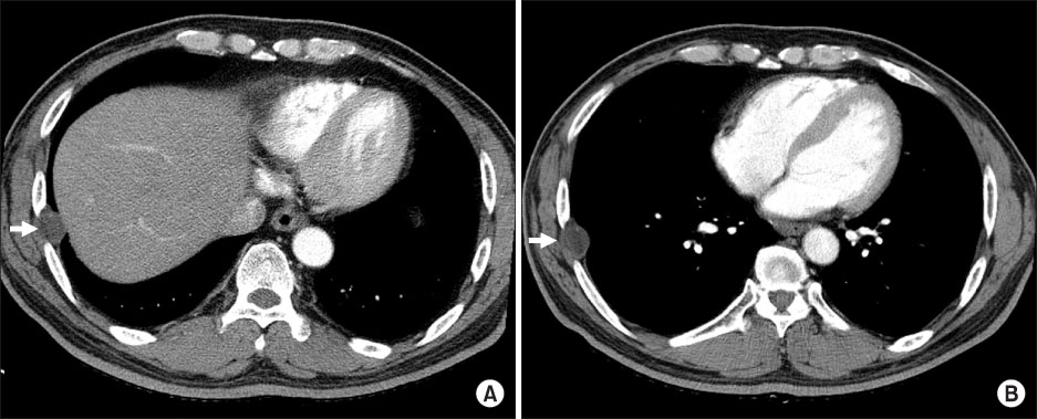
Figure 1 Chest computed tomography scan shows a 2.2×1.9 cm well demarcated pleural based nodule (A, arrow) between the lateral arc of the right seventh and eighth ribs with internal fat density, increasing the size about 4 mm after three months (B, arrow).

Figure 2 Histopathological examination shows the laminated cyst wall (arrow) of Echinococcus with fibrosis in the pleura (A, H&E stain, ×12.5) and the germinal layer (arrow) of Echinococcus (B, H&E stain, ×400).
Reference
-
1. Turgut AT, Altinok T, Topçu S, Koşar U. Local complications of hydatid disease involving thoracic cavity: imaging findings. Eur J Radiol. 2009. 70:49–56.2. Tor M, Atasalihi A, Altuntas N, Sulu E, Senol T, Kir A, et al. Review of cases with cystic hydatid lung disease in a tertiary referral hospital located in an endemic region: a 10 years' experience. Respiration. 2000. 67:539–542.3. Ramos G, Orduña A, García-Yuste M. Hydatid cyst of the lung: diagnosis and treatment. World J Surg. 2001. 25:46–57.4. Ozyurtkan MO, Koçyiğit S, Cakmak M, Ozsoy IE, Balci AE. Case report: secondary pleural hydatidosis. Turkiye Parazitol Derg. 2009. 33:177–178.5. Kireşi DA, Karabacakoğlu A, Odev K, Karaköse S. Uncommon locations of hydatid cysts. Acta Radiol. 2003. 44:622–636.6. Gursoy S, Ucvet A, Tozum H, Erbaycu AE, Kul C, Basok O. Primary intrathoracic extrapulmonary hydatid cysts: analysis of 14 patients with a rare clinical entity. Tex Heart Inst J. 2009. 36:230–233.7. Rakower J, Milwidsk H. Hydatid pleural disease. Am Rev Respir Dis. 1964. 90:623–631.8. Kilani T, El Hammami S. Pulmonary hydatid and other lung parasitic infections. Curr Opin Pulm Med. 2002. 8:218–223.9. Olut AI, Erguven S, Emri S, Ozunlu H, Akay H. Diagnostic value of a dot immunobinding assay for human pulmonary hydatidosis. Korean J Parasitol. 2005. 43:15–18.10. Doğan R, Yūksel M, Cetin G, Sūzer K, Alp M, Kaya S, et al. Surgical treatment of hydatid cysts of the lung: report on 1055 patients. Thorax. 1989. 44:192–199.11. al Karawi MA, Mohamed AR, el Tayeb BO, Yasawy MI. Unintentional percutaneous aspiration of a pleural hydatid cyst. Thorax. 1991. 46:859–860.12. Khan K, Arino J, Hu W, Raposo P, Sears J, Calderon F, et al. Spread of a novel influenza A (H1N1) virus via global airline transportation. N Engl J Med. 2009. 361:212–214.13. MacPherson DW, Gushulak BD, Baine WB, Bala S, Gubbins PO, Holtom P, et al. Population mobility, globalization, and antimicrobial drug resistance. Emerg Infect Dis. 2009. 15:1727–1732.14. Laaksonen S, Solismaa M, Kortet R, Kuusela J, Oksanen A. Vectors and transmission dynamics for Setaria tundra (Filarioidea; Onchocercidae), a parasite of reindeer in Finland. Parasit Vectors. 2009. 2:3.15. Chai JY. Recent trends of parasitic diseases in Korea. Infect Chemother. 2007. 39:Suppl 2. S156–S172.
