Korean J Gastroenterol.
2021 Jan;77(1):35-38. 10.4166/kjg.2020.129.
Hepatic Hydatid Cyst: A Case Report
- Affiliations
-
- 1Division of Gastroenterology, Department of Internal Medicine, Changwon Fatima Hospital, Changwon, Korea
- KMID: 2510774
- DOI: http://doi.org/10.4166/kjg.2020.129
Abstract
- Hydatid cysts are caused by an infestation with larval tapeworms of the genus Echinococcus. The disease is endemic in developing countries but has rarely been reported from immigrant workers in Korea. This paper reports a case of hepatic hydatid cyst in a 27-year-old female. She was referred with abdominal pain that had persisted for the past 2 months. The patient was a foreign worker from Mongolia. The physical examination was unremarkable, and blood tests showed peripheral blood eosinophilia and elevated liver enzymes. Abdominal ultrasonography showed a well-circumscribed cystic mass with septation in the liver. A surgical resection was performed for complete removal. After uncomplicated postoperative recovery, the patient was discharged with albendazole 400 mg twice daily. The hydatid cyst is an important disease that should be considered in the differential diagnosis of cystic lesions in the liver, particularly in those who have lived in endemic areas. A correct early diagnosis based on the typical image findings is important for early treatment before the rupture of the cyst, which is associated with low morbidity and mortality. A current surgical resection combined albendazole are effective treatments for hepatic hydatid cysts, associated with low recurrence rates.
Keyword
Figure
-
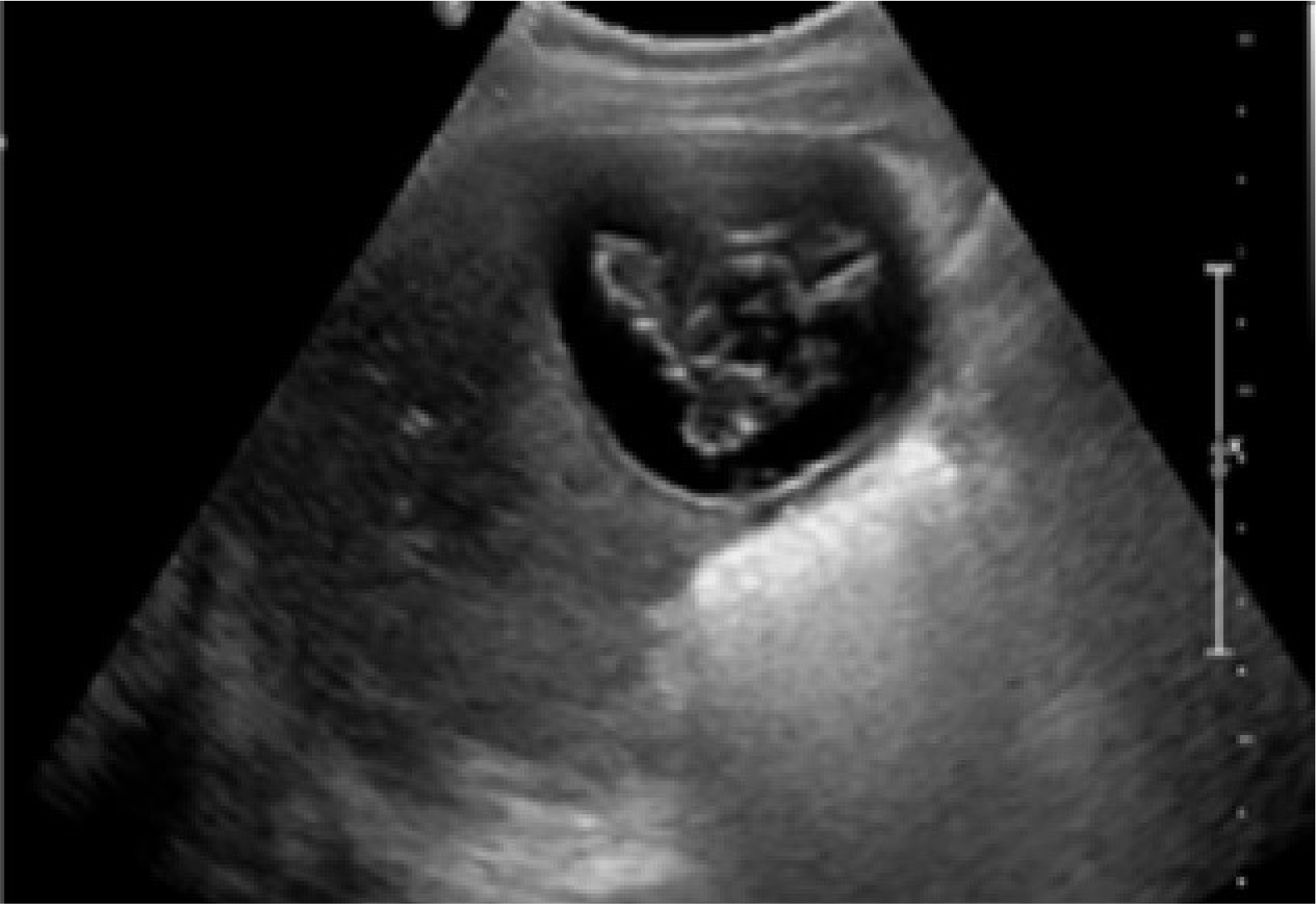
Fig. 1 Abdominal ultrasonography findings, it shows the large cystic lesion has intracystic irregular echogenicities of inner cyst wall infoldings with separation of the hydatid membrane.
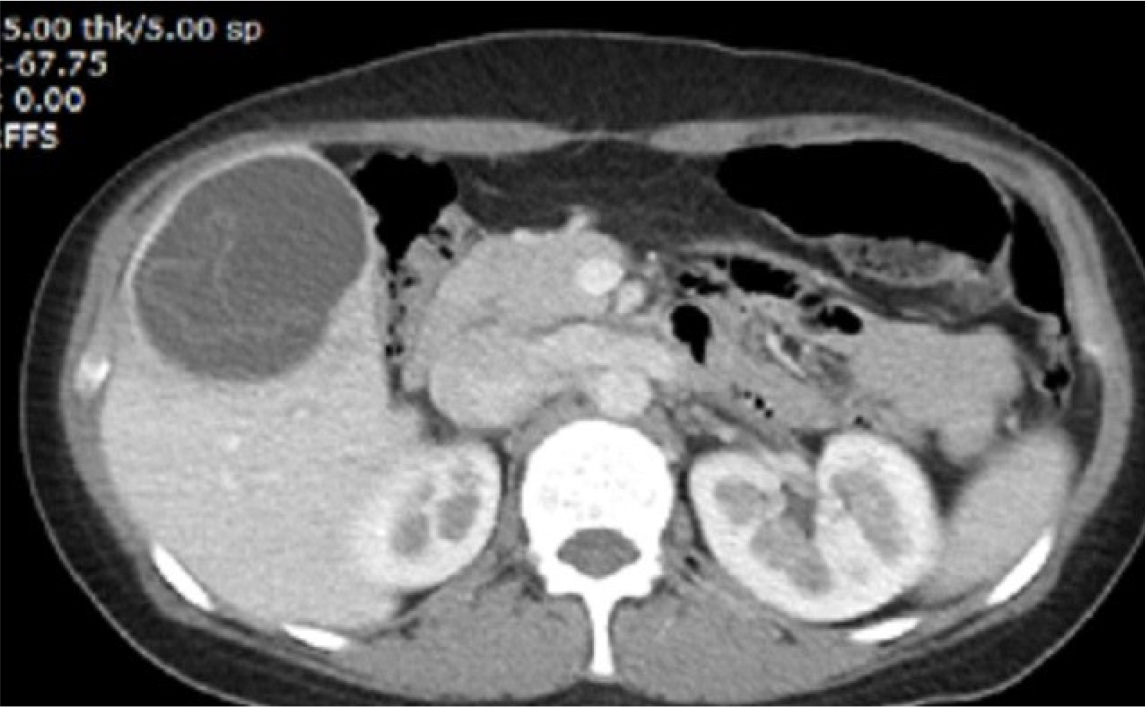
Fig. 2 Abdominal computed tomography scan. It shows a cystic mass with internal septation is present in the right hepatic lobe.
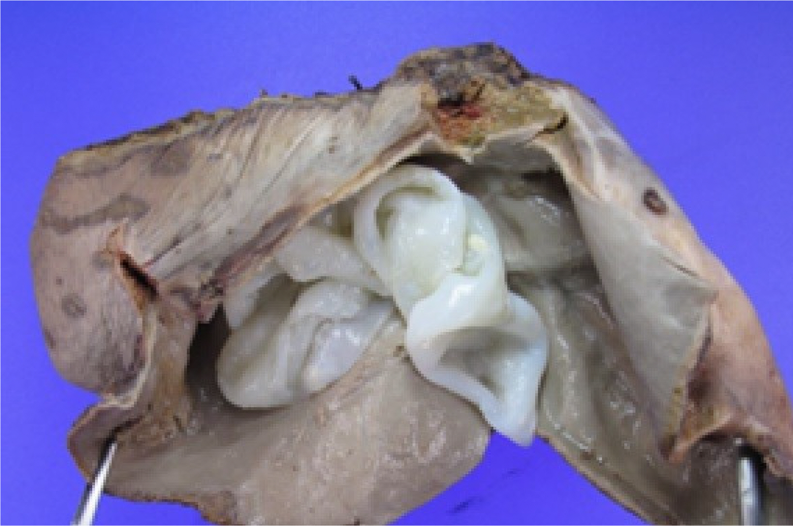
Fig. 3 Glossly, the open mass showed many hydatid cysts after surgery.

Fig. 4 Micropic finding of resected cyst, it shows outer thick laminate menbrane (black arrow) and inner germinal layer is seen (yellow arrow) (H-E stain, ×200).
Reference
-
1. Frider B, Larrieu E, Odriozola M. 1999; Long-term outcome of asymptomatic liver hydatidosis. J Hepatol. 30:228–231. DOI: 10.1016/S0168-8278(99)80066-X. PMID: 10068100.
Article2. Nunnari G, Pinzone MR, Gruttadauria S, et al. 2012; Hepatic echinococcosis: clinical and therapeutic aspects. World J Gastroenterol. 18:1448–1458. DOI: 10.3748/wjg.v18.i13.1448. PMID: 22509076. PMCID: PMC3319940.
Article3. Moro P, Schantz PM. 2009; Echinococcosis: a review. Int J Infect Dis. 13:125–133. DOI: 10.1016/j.ijid.2008.03.037. PMID: 18938096.
Article4. Santivanez S, Garcia HH. 2010; Pulmonary cystic echinococcosis. Curr Opin Pulm Med. 16:257–261. DOI: 10.1097/MCP.0b013e3283386282. PMID: 20216420. PMCID: PMC3362862.
Article5. Jang HJ, Oh MU. 1974; Epizootiological study of echinococcus granulosus( Batsch, 1786) Rudolphi, 1805. in Jeju-do. 1. Incidences of bovine hydatid cyst and its speciation. Korean J Vet. 14:73–76.6. Seo BS, Oh MY, Cho SY. 1975; An echinococcal cyst found inlung of cattle in Cheju-do. Korean J Parasitol. 13:85.7. Park KH, Jung SI, Jang HC, Shin JH. 2009; First successful puncture, aspiration, injection, and re-aspiration of hydatid cyst in the liver presenting with anaphylactic shock in Korea. Yonsei Med J. 50:717–720. DOI: 10.3349/ymj.2009.50.5.717. PMID: 19881979. PMCID: PMC2768250.
Article8. Chai JY, Seo M, Suh KS, Lee SH. 1995; An imported case of hepatic unilocular hydatid disease. Korean J Parasitol. 33:125–130. DOI: 10.3347/kjp.1995.33.2.125. PMID: 7551803.
Article9. Jung BH, Kim TH, Kang JS, et al. 1993; A case of pulmonary and hepatic hydatid cystic disease. Korean J Med. 45:550–556.10. Chae GR, Ann HS, Soo BJ, Chae KM. 1996; A clinical case report of hydatid disease of the liver and lung caused by Echino-coccus granulosus. J Korean Surg Soc. 50:285–290.11. Kim IK, Kim DY, Moon IS, et al. 2004; A case of echinococcal hydatid cyst as misdiagnosed to the bladder cancer. Korean J Urol. 45:1292–1295.12. Ryou JY, Kim KH, Kim SY. 2006; Hydatid cyst of the orbit. J Korean Ophthalmol Soc. 47:484–488. PMID: 29622837. PMCID: PMC5870786.13. Kang MJ, Lee SH, Kim SJ, et al. 2007; A case of multiple intraperitoneal cysts from ruptured hepatic hydatid cysts. Korean J Gastroenterol. 50:203–206. PMID: 17885288.14. Kim IY, Kim SW, Shin HC, Han JK. 2009; US and CT findings of splenic hydatid cyst: a case report. J Korean Soc Ultrasound Med. 28:39–42.15. Kim JM, Choi KW, Lee HJ. 2001; A clinical case report of hydatid cyst of liver. Yeungnam Univ J Med. 18:138–143. DOI: 10.12701/yujm.2001.18.1.138.
Article16. Byun SJ, Moon KC, Suh KS, Han JK, Chai JY. 2010; An imported case of echinococcosis of the liver in a Korean who traveled to Western and Central Europe. Korean J Parasitol. 48:161–165. DOI: 10.3347/kjp.2010.48.2.161. PMID: 20585534. PMCID: PMC2892573.
Article17. Ahn KS, Hong ST, Kang YN, et al. 2012; An imported case of cystic echinococcosis in the liver. Korean J Parasitol. 50:357–360. DOI: 10.3347/kjp.2012.50.4.357. PMID: 23230336. PMCID: PMC3514430.
Article18. Kim AR, Park SJ, Gu MJ, Choi JH, Kim HJ. 2013; Fine needle aspiration cytology of hepatic hydatid cyst: a case study. Korean J Pathol. 47:395–398. DOI: 10.4132/KoreanJPathol.2013.47.4.395. PMID: 24009638. PMCID: PMC3759642.
Article19. Shin DH, Jo HC, Kim JH, et al. 2019; An imported case of disseminated echinococcosis in Korea. Korean J Parasitol. 57:429–434. DOI: 10.3347/kjp.2019.57.4.429. PMID: 31533411. PMCID: PMC6753304.
Article20. Cho E, Shin SS, Kim GE. 2019; Hepatic hydatid disease causing gastric ulcer as a rare complication. J Korean Soc Radiol. 80:987–991. DOI: 10.3348/jksr.2019.80.5.987.
Article
- Full Text Links
-
- Actions
-
Cited
- CITED
-
- Close
- Share
-
- Similar articles
-
- Rupture of Right Hepatic Duct into Hydatid Cyst
- Laparoscopic management of giant hepatic hydatid cyst in a 12-year-old boy: a case report
- An imported case of hepatic unilocular hydatid disease
- Pulmonary Embolism Secondary to Hydatid Cyst of the Right Ventricle: A Case Report
- First Successful Puncture, Aspiration, Injection, and Re-Aspiration of Hydatid Cyst in the Liver Presenting with Anaphylactic Shock in Korea
