A Case of Bronchus-Associated Lymphoid Tissue(BALT) Lymphoma Treated with Lobectomy
- Affiliations
-
- 1Division of Pulmonary, Allergy and Critical Care Medicine of Hallym University Medical Center, Korea. pulmoks@hallym.or.kr
- 2Department of Pathology, Hallym University College of Medicine, Anyang, Korea.
- 3Department of Radiology, Hallym University College of Medicine, Anyang, Korea.
- 4Department of Thorasic and Cardiovascular Surgery Hallym University Sacred Heart Hospital, Hallym University College of Medicine, Anyang, Korea.
Abstract
- The bronchus-asociated lymphoid tissue(BALT) lymphoma is a low-grade primary malignant lymphoma that originates from bronchus associated lymphoid tissue. A 67-year-old woman was admitted for evaluation of cough, sputum, rhinorrhea which had persisted for one month. Physical examination showed decreased breathing sound on the left upper lung field. High resolution chest computed tomography demonstrated consolidation which showed air-bronchogram and surrounding ground glass opacity in left upper lobe. These findings implicated inactive tuberculosis, organizing pneumonia, or bronchiolo-alveolar carcinoma. The histologic findings from percutaneous needle aspiration biopsy revealed aggregated atypical small lymphoid cells with lymphoepithelial lesions. With immunohistochemical staining, the atypical lymphoid cells reacted positively with CD 20 antibody and negatively with CD 3 antibody. Thus, we could diagnosed her as a patient with BALT lymphoma. After left upper lobectomy, she has been well without recurrence of the disease for 14 months. In this country of Republic of Korea, it was the 1st case of BALT lymphoma surgically treated when histological diagnosis had been done. Based on this case, we wanted to demonstrate the importance of early histological diagnosis and treatment of BALT lymphoma.
MeSH Terms
Figure
-
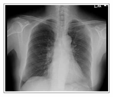
Figure 1A Consolidation combined with linear opacities was noted on left upper lobe.

Figure 1B HRCT demonstrated consolidation which showed air-bronchogram and surrounding ground glass opacity in left upper lobe and the lesion was mainly adjacent to pleura and fissure.

Figure 2 In gross appearance after operation, lesion demonstrated an irregular diffuse consolidation with a solid tan-colored cut surface abutting visceral pleura.

Figure 3 On 12 months After lobectomy, whole body PET CT did not demonstrate any evidence of local recurrence or distant metastasis of BALT lymphoma.

Figure 4 (A) Atypical lymphocytes with interstitial multinodular infiltration pattern and multiple foci of perivascular lymphoid infiltration ("lymphotic trackings", arrows) were seen. (H&E, ×100) (B) The tumor consisted of small lymphocytes which showed clear abundant cytoplasm. They grew in monotonous sheets and infiltrated to respiratory epithelial cells("lymphoepithelial lesion", arrows). (H&E, ×200) (C) Atypical lymphocytes reacted with CD 20 antibody positively (B cell maker). (immunohistochemistry, ×200) (D) Epithelial cells in the lymphoepithelial lesions reacted with cytokeratin positively . (immunohistochemistry, ×200)
Reference
-
1. Freeman C, Berg JW, Cutler SJ. Occurrence and prognosis of extranodal lymphomas. Cancer. 1972. 29:252–260.2. Cadranel J, Wislez M, Antoine M. Primary pulmonary lymphoma. Eur Respir J. 2002. 20:750–762.3. Michael CW, Richardson PH, Boudreaux CW. Pulmonary lymphoma of the mucosa-associated lymphoid tissue type: report of a case with cytological, histological, immunophenotypical correlation, and review of the literature. Ann Diagn Pathol. 2005. 9:148–152.4. Ahmed S, Kussick SJ, Siddiqui AK, Bhuiya TA, Khan A, Sarewitz S, et al. Bronchial-associated lymphoid tissue lymphoma: a clinical study of a rare disease. Eur J Cancer. 2004. 40:1320–1326.5. Montes M, Tomasi TB Jr, Noehren TH, Culver GJ. Lymphoid interstitial pneumonia with monoclonal gammopathy. Am Rev Respir Dis. 1968. 98:277–280.6. Cordier JF, Cellier CC, Vincent M, Loire R, Creyssel R, Brune J. Monoclonal gammopathies in chest disease. Thrax. 1985. 40:629–630.7. Lee DK, Im JG, Lee KS, Lee JS, Seo JB, Goo JM, et al. B-cell lymphoma of bronchus-associated lymphoid tissue(BALT): CT features in 10 patients. J Comput Assist Tomogr. 2000. 24:30–34.8. Takamori M, Noma S, Kobashi Y, Inoue T, Gohma I, Mino M, et al. CT findings of BALTOMA. Radiat Med. 1999. 17:349–354.9. Lee SM, Yoon HI, Choi SH, Hwangbo B, Yoo CG, Lee CT, et al. Cases of the pulmonary malignant lymphoma of the Bronchus-Associated Lymphoid Tissue (BALT). Tuberc and Respir Dis. 1999. 47:681–687.10. Nicholson AG, Wotherspoon AC, Diss TC, Butcher DN, Sheppard MN, Isaacson PG, et al. Pulmonary B-cell non-Hodgkin's lymphomas. The value of immunohistochemistry and gene analysis in diagnosis. Histopathology. 1995. 26:395–403.11. Thieblemont C, Berger F, Dumontet C, Moullet I, Bouafia F, Felman P, et al. Mucosa-associated lymphoid tissue lymphoma is a disseminated disease in one third of 158 patients analyzed. Blood. 2000. 95:802–806.
- Full Text Links
-
- Actions
-
Cited
- CITED
-
- Close
- Share
-
- Similar articles
-
- Cases of the Pulmonary Malignant Lymphoma of the Bronchus-Associated Lymphoid Tissue (BALT)
- Mucosa-Associated Lymphoid Tissue Lymphoma of the Esophagus Coexistent with Bronchus-Associated Lymphoid Tissue Lymphoma of the Lung
- A case of bronchus-associated lymphoid tissue (BALT) lymphoma in the patient with rheumatoid arthritis
- Radiation Therapy for Bronchial Associated Lymphoid Tissue (BALT) Lymphoma : A case report
- Higher Microbial Abundance and Diversity in Bronchus-Associated Lymphoid Tissue Lymphomas Than in Non-cancerous Lung Tissues
