Restor Dent Endod.
2014 Nov;39(4):276-281. 10.5395/rde.2014.39.4.276.
Cutting efficiency of apical preparation using ultrasonic tips with microprojections: confocal laser scanning microscopy study
- Affiliations
-
- 1Department of Conservative Dentistry, School of Dentistry, Pusan National University, Dental Research Institute, Yangsan, Korea. golddent@pusan.ac.kr
- 2Department of Conservative Dentistry, School of Dentistry, Daejeon Hospital, WonKwang University, Daejeon, Korea.
- 3Department of Conservative Dentistry, School of Dentistry, Seoul National University, Dental Research Institute, Seoul, Korea.
- KMID: 2316908
- DOI: http://doi.org/10.5395/rde.2014.39.4.276
Abstract
OBJECTIVES
The purpose of this study was to compare the cutting efficiency of a newly developed microprojection tip and a diamond-coated tip under two different engine powers.
MATERIALS AND METHODS
The apical 3-mm of each root was resected, and root-end preparation was performed with upward and downward pressure using one of the ultrasonic tips, KIS-1D (Obtura Spartan) or JT-5B (B&L Biotech Ltd.). The ultrasonic engine was set to power-1 or -4. Forty teeth were randomly divided into four groups: K1 (KIS-1D / Power-1), J1 (JT-5B / Power-1), K4 (KIS-1D / Power-4), and J4 (JT-5B / Power-4). The total time required for root-end preparation was recorded. All teeth were resected and the apical parts were evaluated for the number and length of cracks using a confocal scanning micrscope. The size of the root-end cavity and the width of the remaining dentin were recorded. The data were statistically analyzed using two-way analysis of variance and a Mann-Whitney test.
RESULTS
There was no significant difference in the time required between the instrument groups, but the power-4 groups showed reduced preparation time for both instrument groups (p < 0.05). The K4 and J4 groups with a power-4 showed a significantly higher crack formation and a longer crack irrespective of the instruments. There was no significant difference in the remaining dentin thickness or any of the parameters after preparation.
CONCLUSIONS
Ultrasonic tips with microprojections would be an option to substitute for the conventional ultrasonic tips with a diamond coating with the same clinical efficiency.
Keyword
Figure
-
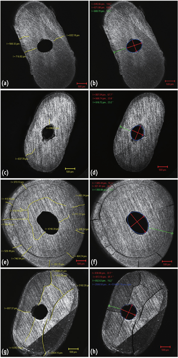
Figure 1 The crack length and shape measurement after cavity preparation. (a and b) Group K1 by KIS-1D tip and power 1; (c and d) group J1 by JT-5B tip and power 1; (e and f) group K4 by KIS-1D tip and power 4; (g and h) group J4 by JT-5B tip and power 4.
Cited by 1 articles
-
Questioning the spot light on Hi-tech endodontics
Jojo Kottoor, Denzil Albuquerque
Restor Dent Endod. 2016;41(1):80-82. doi: 10.5395/rde.2016.41.1.80.
Reference
-
1. Sjogren U, Hagglund B, Sundqvist G, Wing K. Factors affecting the long-term results of endodontic treatment. J Endod. 1990; 16:498–504.
Article2. Plotino G, Pameijer CH, Grande NM, Somma F. Ultrasonics in endodontics: a review of the literature. J Endod. 2007; 33:81–95.
Article3. Engel TK, Steiman HR. Preliminary investigation of ultrasonic root end preparation. J Endod. 1995; 21:443–445.
Article4. Gondim E Jr, Gomes BP, Ferraz CC, Teixeira FB, Souza-Filho FJ. Effect of sonic and ultrasonic retrograde cavity preparation on the integrity of root apices of freshly extracted human teeth: scanning electron microscopy analysis. J Endod. 2002; 28:646–650.
Article5. Tidmarsh BG, Arrowsmith MG. Dentinal tubules at the root ends of apicected teeth: a scanning electron microscopic study. Int Endod J. 1989; 22:184–189.
Article6. Gutmann JL, Harrison JW. Posterior endodontic surgery: anatomical considerations and clinical techniques. Int Endod J. 1985; 18:8–34.
Article7. Gutmann JL, Saunders WP, Nguyen L, Guo IY, Saunders EM. Ultrasonic root-end preparation. Part 1. SEM analysis. Int Endod J. 1994; 27:318–324.
Article8. Bertrand G, Festal F, Barailly R. Use of ultrasound in apico-ectomy. Quintessence Int. 1976; 7:9–12.9. Lin YH, Mickel AK, Jones JJ, Montagnese TA, González AF. Evaluation of cutting efficiency of ultrasonic tips used in orthograde endodontic treatment. J Endod. 2006; 32:359–361.
Article10. Brent PD, Morgan LA, Marshall JG, Baumgartner JC. Evaluation of diamond-coated ultrasonic instruments for root-end preparation. J Endod. 1999; 25:672–675.
Article11. Saunders WP, Saunders EM, Gutmann JL. Ultrasonic root-end preparation, Part 2. Microleakage of EBA root-end fillings. Int Endod J. 1994; 27:325–329.
Article12. Layton CA, Marshall JG, Morgan LA, Baumgartner JC. Evaluation of cracks associated with ultrasonic root-end preparation. J Endod. 1996; 22:157–160.
Article13. Abedi RH, Van Mierlo BL, Wilder-Smith P, Torabinejad M. Effects of ultrasonic root-end cavity preparation on the root apex. Oral Surg Oral Med Oral Pathol Oral Radiol Endod. 1995; 80:207–213.
Article14. Waplington M, Lumley PJ, Walmsley AD. Incidence of root face alteration after ultrasonic retrograde cavity preparation. Oral Surg Oral Med Oral Pathol Oral Radiol Endod. 1997; 83:387–392.
Article15. Frank RJ, Antrim DD, Bakland LK. Effect of retrograde cavity preparations on root apexes. Endod Dent Traumatol. 1996; 12:100–103.
Article16. von Arx T, Walker WA 3rd. Microsurgical instruments for root-end cavity preparation following apicoectomy: a literature review. Endod Dent Traumatol. 2000; 16:47–62.
Article17. Gray GJ, Hatton JF, Holtzmann DJ, Jenkins DB, Nielsen CJ. Quality of root-end preparations using ultrasonic and rotary instrumentation in cadavers. J Endod. 2000; 26:281–283.
Article18. Calzonetti KJ, Iwanowski T, Komorowski R, Friedman S. Ultrasonic root end cavity preparation assessed by an in situ impression technique. Oral Surg Oral Med Oral Pathol Oral Radiol Endod. 1998; 85:210–215.
Article19. Khabbaz MG, Kerezoudis NP, Aroni E, Tsatsas V. Evaluation of different methods for the root-end cavity preparation. Oral Surg Oral Med Oral Pathol Oral Radiol Endod. 2004; 98:237–242.
Article20. Mehlhaff DS, Marshall JG, Baumgartner JC. Comparison of ultrasonic and high-speed-bur root-end preparations using bilaterally matched teeth. J Endod. 1997; 23:448–452.
Article21. Engel TK, Steiman HR. Preliminary investigation of ultrasonic root end preparation. J Endod. 1995; 21:443–445.
Article22. Beling KL, Marshall JG, Morgan LA, Baumgartner JC. Evaluation for cracks associated with ultrasonic root-end preparation of gutta-percha filled canals. J Endod. 1997; 23:323–326.
Article23. Taschieri S, Testori T, Francetti L, Del Fabbro M. Effects of ultrasonic root end preparation on resected root surfaces: SEM evaluation. Oral Surg Oral Med Oral Pathol Oral Radiol Endod. 2004; 98:611–618.
Article
- Full Text Links
-
- Actions
-
Cited
- CITED
-
- Close
- Share
-
- Similar articles
-
- Effect of three different irrigation solutions applied by passive ultrasonic irrigation
- The Effects of a Er:YAG Laser on Machined, Sand-Blasted and Acid-Etched, and Resorbable Blast Media Titanium Surfaces Using Confocal Microscopy and Scanning Electron Microscopy
- Intravital Laser-scanning Two-photon and Confocal Microscopy for Biomedical Research
- Application of Autofluorescence for Confocal Microscopy to Aid in Archaeoparasitological Analyses
- An experimental study of cutting efficiency of air-driven diamond burs on human tooth
