Restor Dent Endod.
2014 Feb;39(1):39-44.
Cytotoxicity of newly developed pozzolan cement and other root-end filling materials on human periodontal ligament cell
- Affiliations
-
- 1Department of Conservative Dentistry Gangnam Severance Dental Hospital, Yonsei University College of Dentistry, Seoul, Korea.
- 2Department of Conservative Dentistry, Yonsei University College of Dentistry, Seoul, Korea.
- 3Microscope Center, Department of Conservative Dentistry and Oral Science Research Center, Yonsei University College of Dentistry, Seoul, Korea. andyendo@yuhs.ac
Abstract
OBJECTIVES
The purpose of this study was to evaluate in vitro cytotoxicity of the pozzolan cement and other root-end filling materials using human periodontal ligament cell.
MATERIALS AND METHODS
Endocem (Maruchi), white ProRoot MTA (Dentsply), white Angelus MTA (Angelus), and Super EBA (Bosworth Co.) were tested after set completely in an incubator at 37degrees C for 7 days, Endocem was tested in two ways: 1) immediately after mixing (fresh specimens) and 2) after setting completely like other experimental materials. The methods for assessment included light microscopic examination, cell counting and WST-1 assay on human periodontal ligament cell.
RESULTS
In the results of microscopic examination and cell counting, Super EBA showed significantly lower viable cell than any other groups (p < 0.05). As the results of WST-1 assay, compared with untreated control group, there was no significant cell viability of the Endocem group. However, the fresh mixed Endocem group had significantly less cell viability. The cells exposed to ProRoot MTA and Angelus MTA showed the highest viability, whereas the cells exposed to Super EBA displayed the lowest viability (p < 0.05).
CONCLUSIONS
The cytotoxicity of the pozzolan cement (Endocem) was comparable with ProRoot MTA and Angelus MTA. Considering the difficult manipulation and long setting time of ProRoot MTA and Angelus MTA, Endocem can be used as the alternative of retrofilling material.
Figure
-
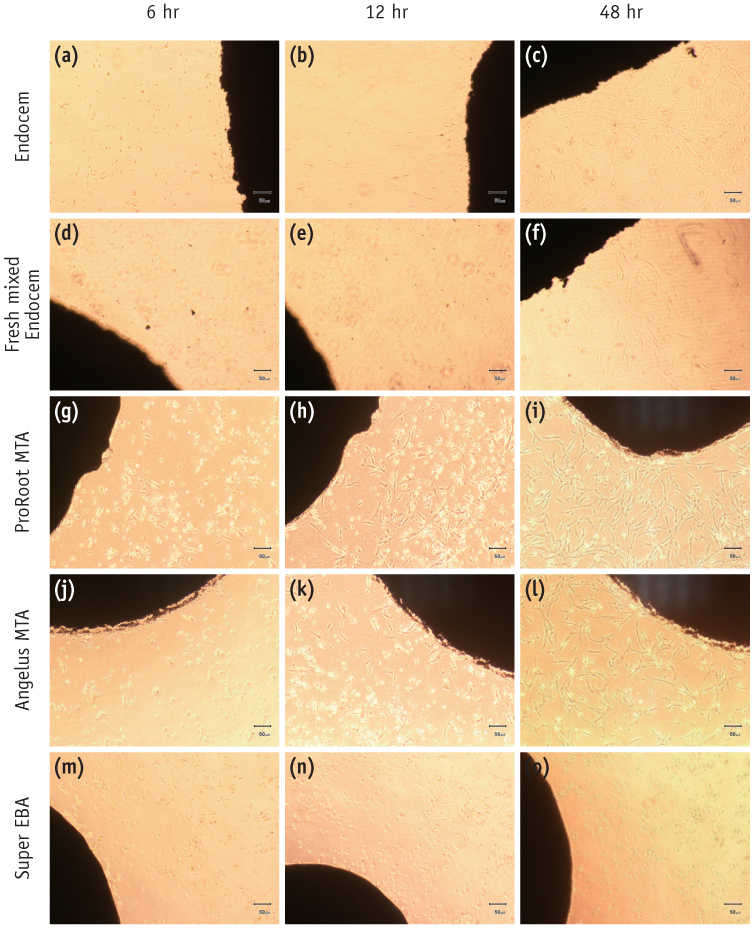
Figure 1 Morphological changes of periodontal ligament cells in contact with each experimental material. (a - c) Endocem; (d - f) fresh mixed Endocem; (g - i) ProRoot MTA; (j - l) Angelus MTA; (m - o) Super EBA at 6, 12, and 48 hours.
Reference
-
1. Torabinejad M, Hong CU, Pitt Ford TR, Kaiyawasam SP. Tissue reaction to implanted super-EBA and mineral trioxide aggregate in the mandible of guinea pigs: a preliminary report. J Endod. 1995; 21:569–571.
Article2. Al-Sa'eed OR, Al-Hiyasat AS, Darmani H. The effects of six root-end filling materials and their leachable components on cell viability. J Endod. 2008; 34:1410–1414.3. Bondra DL, Hartwell GR, MacPherson MG, Portell FR. Leakage in vitro with IRM, high copper amalgam, and EBA cement as retrofilling materials. J Endod. 1989; 15:157–160.
Article4. Samara A, Sarri Y, Stravopodis D, Tzanetakis GN, Kontakiotis EG, Anastasiadou E. A comparative study of the effects of three root-end filling materials on proliferation and adherence of human periodontal ligament fibroblasts. J Endod. 2011; 37:865–870.
Article5. Bodrumlu E. Biocompatibility of retrograde root filling materials: a review. Aust Endod J. 2008; 34:30–35.
Article6. Asgary S, Eghbal MJ, Parirokh M, Ghoddusi J, Kheirieh S, Brink F. Comparison of mineral trioxide aggregate's composition with Portland cements and a new endodontic cement. J Endod. 2009; 35:243–250.
Article7. Dorn SO, Gartner AH. Retrograde filling materials: A retrospective success-failure study of amalgam, EBA, and IRM. J Endod. 1990; 16:391–393.
Article8. Torabinejad M, Hong CU, McDonald F, Pitt Ford TR. Physical and chemical properties of a new root-end filling material. J Endod. 1995; 21:349–353.
Article9. Torabinejad M, Hong CU, Lee SJ, Monsef M, Pitt Ford TR. Investigation of mineral trioxide aggregate for root-end filling in dogs. J Endod. 1995; 21:603–608.
Article10. Rubinstein RA, Kim S. Long-term follow-up of cases considered healed one year after apical microsurgery. J Endod. 2002; 28:378–383.
Article11. Taschieri S, Del Fabbro M, Testori T, Weinstein R. Endoscopic periradicular surgery: a prospective clinical study. Br J Oral Maxillofac Surg. 2007; 45:242–244.
Article12. Enkel B, Dupas C, Armengol V, Akpe Adou J, Bosco J, Daculsi G, Jean A, Laboux O, LeGeros RZ, Weiss P. Bioactive materials in endodontics. Expert Rev Med Devices. 2008; 5:475–494.
Article13. Parirokh M, Torabinejad M. Mineral trioxide aggregate: a comprehensive literature review-Part I: chemical, physical, and antibacterial properties. J Endod. 2010; 36:16–27.
Article14. Parirokh M, Torabinejad M. Mineral trioxide aggregate: a comprehensive literature review-Part III: Clinical applications, drawbacks, and mechanism of action. J Endod. 2010; 36:400–413.
Article15. Torabinejad M, Parirokh M. Mineral trioxide aggregate: a comprehensive literature review-part II: leakage and biocompatibility investigations. J Endod. 2010; 36:190–202.
Article16. Song M, Kim SG, Shin SJ, Kim HC, Kim E. The influence of bone tissue deficiency on the outcome of endodontic microsurgery: a prospective study. J Endod. 2013; 39:1341–1345.
Article17. Karimjee CK, Koka S, Rallis DM, Gound TG. Cellular toxicity of mineral trioxide aggregate mixed with an alternative delivery vehicle. Oral Surg Oral Med Oral Pathol Oral Radiol Endod. 2006; 102:e115–e120.
Article18. Huang FM, Chang YC. Cytotoxicity of resin-based restorative materials on human pulp cell cultures. Oral Surg Oral Med Oral Pathol Oral Radiol Endod. 2002; 94:361–365.
Article19. Geurtsen W, Lehmann F, Spahl W, Leyhausen G. Cytotoxicity of 35 dental resin composite monomers/additives in permanent 3T3 and three human primary fibroblast cultures. J Biomed Mater Res. 1998; 41:474–480.
Article20. Pérez AL, Spears R, Gutmann JL, Opperman LA. Osteoblasts and MG-63 osteosarcoma cells behave differently when in contact with ProRoot MTA and White MTA. Int Endod J. 2003; 36:564–570.
Article21. Abdullah D, Ford TR, Papaioannou S, Nicholson J, McDonald F. An evaluation of accelerated Portland cement as a restorative material. Biomaterials. 2002; 23:4001–4010.
Article22. Balto HA. Attachment and morphological behavior of human periodontal ligament fibroblasts to mineral trioxide aggregate: a scanning electron microscope study. J Endod. 2004; 30:25–29.
Article23. Zhu Q, Haglund R, Safavi KE, Spangberg LS. Adhesion of human osteoblasts on root-end filling materials. J Endod. 2000; 26:404–406.
Article24. Ishiyama M, Tominaga H, Shiga M, Sasamoto K, Ohkura Y, Ueno K. A combined assay of cell viability and in vitro cytotoxicity with a highly water-soluble tetrazolium salt, neutral red and crystal violet. Biol Pharm Bull. 1996; 19:1518–1520.
Article25. Ishiyama M, Shiga M, Sasamoto K, Mizoguchi M, He PG. A new sulfonated tetrazolium salt that produces a highly water-soluble formazan dye. Chem Pharm Bull (Tokyo). 1993; 41:1118–1122.
Article26. De Deus G, Ximenes R, Gurgel-Filho ED, Plotkowski MC, Coutinho-Filho T. Cytotoxicity of MTA and Portland cement on human ECV 304 endothelial cells. Int Endod J. 2005; 38:604–609.
Article27. Saidon J, He J, Zhu Q, Safavi K, Spångberg LS. Cell and tissue reactions to mineral trioxide aggregate and Portland cement. Oral Surg Oral Med Oral Pathol Oral Radiol Endod. 2003; 95:483–489.
Article28. Shin SJ. In vitro studies addressing celluar mechanisms underlying the bone and dentin inductive property of mineral trioxide aggregate (MTA). University of Pennsylvania;2004. 75. Master thesis in oral Biology.29. Lin CP, Chen YJ, Lee YL, Wang JS, Chang MC, Lan WH, Chang HH, Chao WM, Tai TF, Lee MY, Lin BR, Jeng JH. Effects of root-end filling materials and eugenol on mitochondrial dehydrogenase activity and cytotoxicity to human periodontal ligament fibroblasts. J Biomed Mater Res B Appl Biomater. 2004; 71:429–440.
Article30. Bonson S, Jeansonne BG, Lallier TE. Root-end filling materials alter fibroblast differentiation. J Dent Res. 2004; 83:408–413.
Article31. Moghaddame-Jafari S, Mantellini MG, Botero TM, McDonald NJ, Nör JE. Effect of ProRoot MTA on pulp cell apoptosis and proliferation in vitro. J Endod. 2005; 31:387–391.
- Full Text Links
-
- Actions
-
Cited
- CITED
-
- Close
- Share
-
- Similar articles
-
- Effects of four novel root-end filling materials on the viability of periodontal ligament fibroblasts
- Biocompatibility of root-end filling materials: recent update
- Evaluation of the radiopacity and cytotoxicity of resinous root canal sealers
- Biocompatibility of bioaggregate cement on human pulp and periodontal ligament (PDL) derived cells
- Washout resistance of fast-setting pozzolan cement under various root canal irrigants
