Wilms' Tumor in a Horseshoe Kidney
- Affiliations
-
- 1Department of Urology, Kangbuk Samsung Hospital, Sungkyunkwan University School of Medicine, Seoul, Korea. hjae.park@samsung.com
Abstract
- The incidence of horseshoe kidney is about 1 in 400 cases. The presence of Wilms' tumor with a horseshoe kidney is unusual, and the occurrence of Wilms' tumor in a horseshoe kidney is estimated at 0.4 to 0.9% of all Wilms' tumors. We report the case of a 5-year-old boy who presented with a stage IV Wilms' tumor in a horseshoe kidney. The patient was treated with preoperative chemotherapy followed by surgical resection and adjuvant chemotherapy. This case illustrates the role of preoperative chemotherapy for preserving renal function and aims to highlight the multimodality treatment of Wilms' tumor.
Figure
-
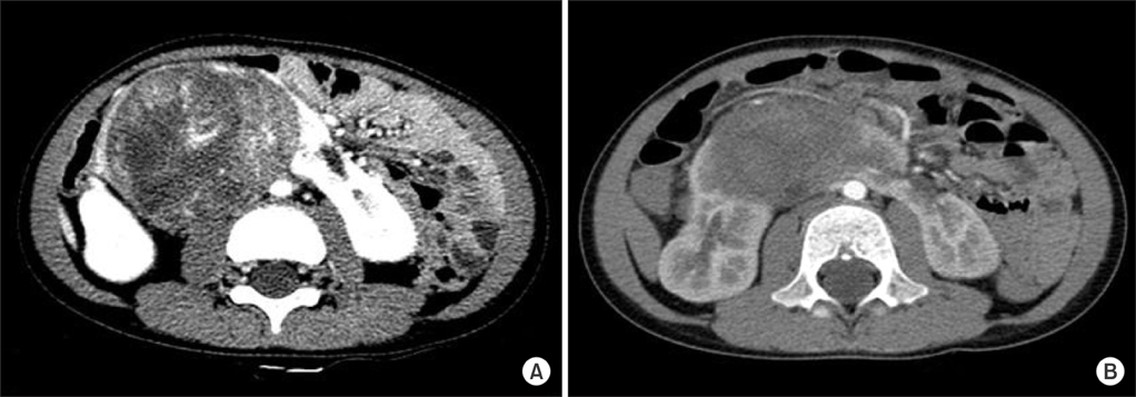
FIG. 1 (A) Computed tomography (CT) scan of the abdomen showing a large heterogenous mass arising from the isthmus of a horseshoe kidney with a parenchymal band extending across the midline (before chemotherapy). (B) CT scan of the abdomen after neoadjuvant chemotherapy showing a decrease in tumor size and central necrotic changes.
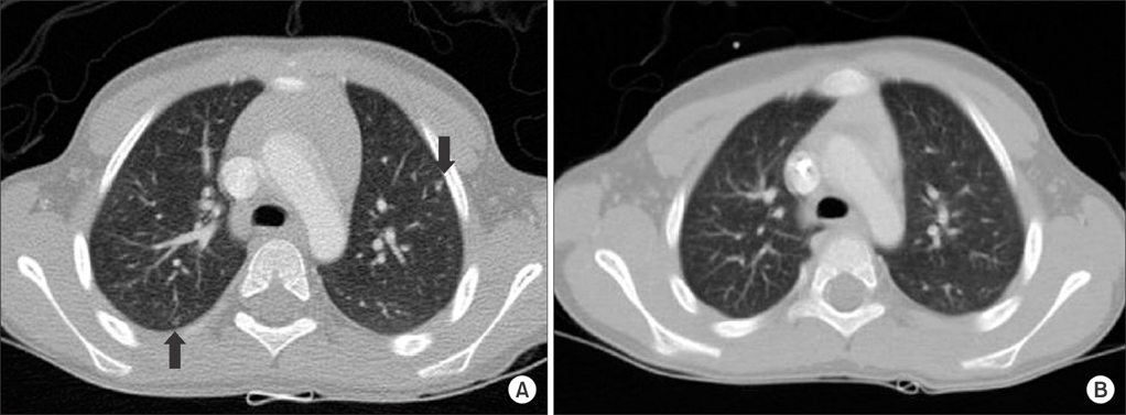
FIG. 2 (A) Computed tomography scan of the chest before chemotherapy showing metastatic small nodules (black arrows). (B) After chemotherapy, the metastatic small nodules disappeared.
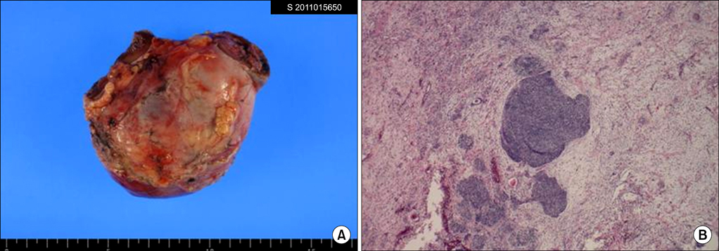
FIG. 3 Histopathological findings. (A) Grossly, the excised mass originated from the isthmus of the horseshoe kidney and surrounded the lower poles of both kidneys. (B) Microscopic findings. Typical Wilms' tumor with blastemal, epithelial, and stromal components (H&E, ×40).
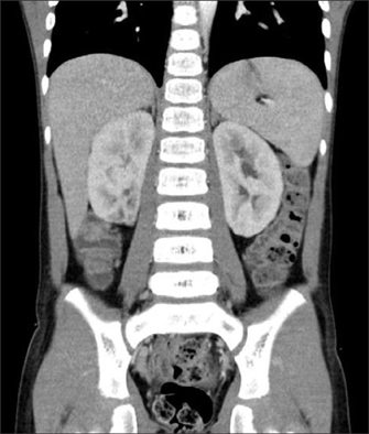
FIG. 4 Follow-up computed tomography scan of the abdomen (coronal view) after the completion of adjuvant chemotherapy in 25 weeks.
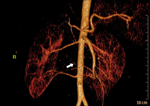
FIG. 5 Reconstructed computed tomography angiogram showing multiple renal arteries and aberrant vascular supplies (white arrow) to the isthmus and tumor.
Reference
-
1. Petruzzi MJ, Green DM. Wilms' tumor. Pediatr Clin North Am. 1997. 44:939–952.2. Mesrobian HG, Kelalis PP, Hrabovsky E, Othersen HB Jr, deLorimier A, Nesmith B. Wilms tumor in horseshoe kidneys: a report from the National Wilms Tumor Study. J Urol. 1985. 133:1002–1003.3. van der Poel HG, Feitz WF, Bokkerink J, Staak FV, de Vries JD. Wilms tumor with teratomatous cysts in a horseshoe kidney: a diagnostic pitfall. J Urol. 1997. 157:1837–1838.4. Neville H, Ritchey ML, Shamberger RC, Haase G, Perlman S, Yoshioka T. The occurrence of Wilms tumor in horseshoe kidneys: a report from the National Wilms Tumor Study Group (NWTSG). J Pediatr Surg. 2002. 37:1134–1137.5. Park JS, Baek SH, Lim JK, Choi JH, Hwa JS, Chung KH. A case of Wilms' tumor arising in horseshoe kidney. Korean J Urol. 2001. 42:550–552.6. Davidoff AM. Wilms' tumor. Curr Opin Pediatr. 2009. 21:357–364.7. Ahmed HU, Arya M, Tsiouris A, Sellaturay SV, Shergill IS, Duffy PG, et al. An update on the management of Wilms' tumour. Eur J Surg Oncol. 2007. 33:824–831.8. Kolln CP, Boatman DL, Schmidt JD, Flocks RH. Horseshoe kidney: a review of 105 patients. J Urol. 1972. 107:203–204.9. Trulock TS, Ricketts RR, Verras A, Kim TH, Hawkins HK, Woodard JR. Wilms tumor arising in horseshoe kidney. Urology. 1985. 25:306–309.10. Pappis CH, Moussatos GH, Constantinides CG, Kairis M. Bilateral nephroblastoma in a horseshoe kidney. J Pediatr Surg. 1979. 14:483–484.
