Obstet Gynecol Sci.
2015 Mar;58(2):150-156. 10.5468/ogs.2015.58.2.150.
Innervation in women with uterine myoma and adenomyosis
- Affiliations
-
- 1Department of Obstetrics and Gynecology, Ilsan Paik Hospital, Inje University College of Medicine, Goyang, Korea. camanbal@gmail.com
- 2Department of Pathology, Ilsan Paik Hospital, Inje University College of Medicine, Goyang, Korea.
- KMID: 2314039
- DOI: http://doi.org/10.5468/ogs.2015.58.2.150
Abstract
OBJECTIVE
To determine if neurofilament (NF) is expressed in the endometrium and the lesions of myomas and adenomyosis, and to determine their correlation.
METHODS
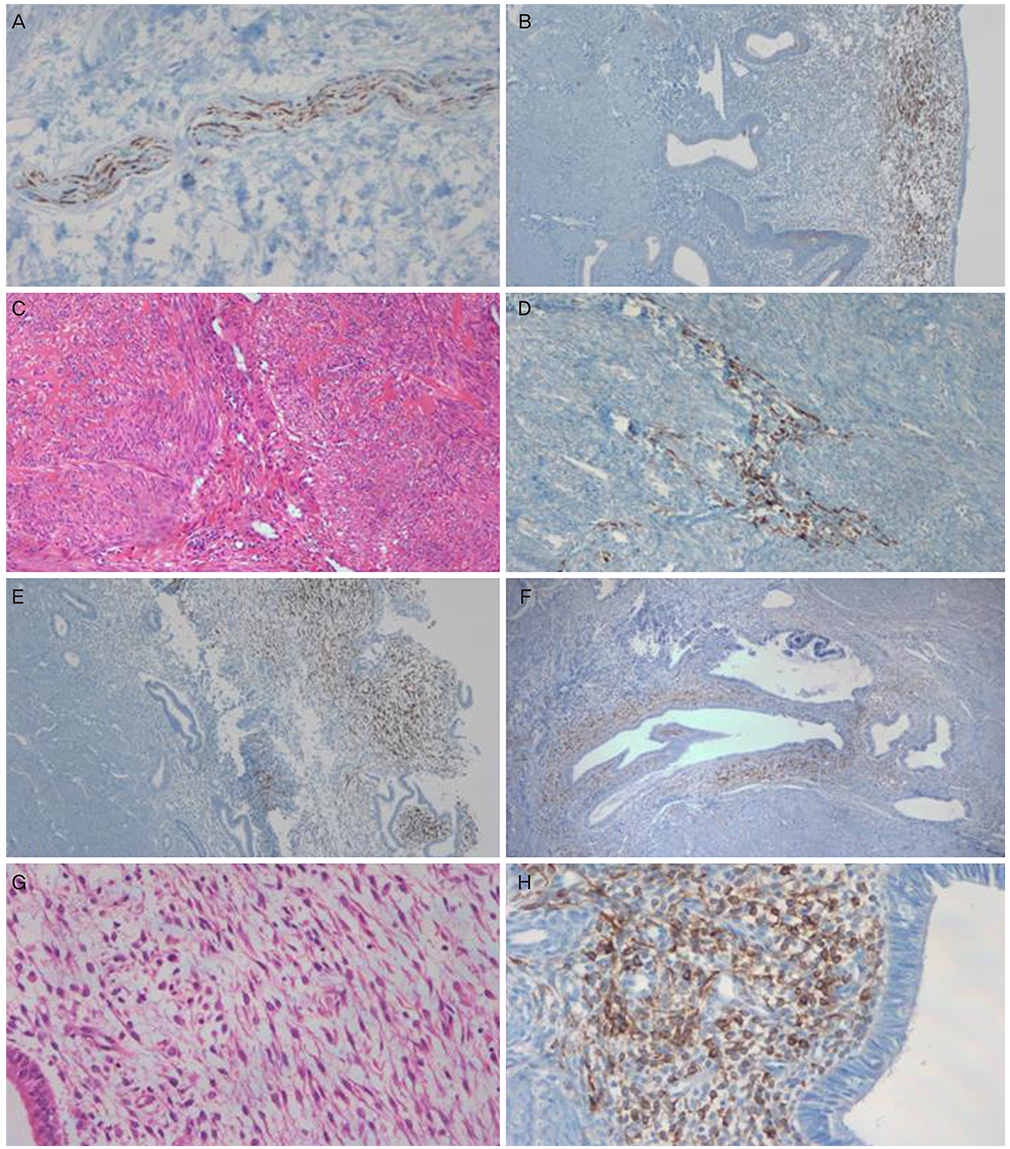
Histologic sections were prepared from hysterectomies performed on women with adenomyosis (n=21), uterine myoma (n=31), and carcinoma in situ of the uterine cervix. Full-thickness uterine paraffin blocks, which included the endometrium and myometrium histologic sections, were stained immunohistochemically using the antibodies for monoclonal mouse antihuman NF protein.
RESULTS
NF-positive cells were found in the endometrium and myometrium in 11 women with myoma and in 7 with adenomyosis, but not in patients with carcinoma in situ of uterine cervix, although the difference was statistically not significant. There was no significant difference between the existence of NF-positive cells and menstrual pain or phases. The NF-positive nerve fibers were in direct contact with the lesions in nine cases (29.0%) of myoma and in five cases (23.8%) of adenomyosis. It was analyzed if there was a statistical significance between the existence of NF positive cells in the endometrium and the expression of NF-positive cells in the uterine myoma/adenomyosis lesions. When NF-positive cell were detected in the myoma lesions, the incidence of NF-positive nerve cells in the eutopic endometrium was significantly high. When NF-positive cell were detected in the basal layer, the incidence of NF-positive nerve cells in the myoma lesions and adenomyosis lesions was significantly high.
CONCLUSION
We assume that NF-positive cells in the endometrium and the myoma and adenomyosis lesions might play a role in pathogenesis. Therefore, more studies may be needed on the mechanisms of nerve fiber growth in estrogen-dependent diseases.
Keyword
MeSH Terms
Figure
Reference
-
1. Krantz KE. Innervation of the human uterus. Ann N Y Acad Sci. 1959; 75:770–784.2. Tokushige N, Markham R, Russell P, Fraser IS. High density of small nerve fibres in the functional layer of the endometrium in women with endometriosis. Hum Reprod. 2006; 21:782–787.3. Atwal G, du Plessis D, Armstrong G, Slade R, Quinn M. Uterine innervation after hysterectomy for chronic pelvic pain with, and without, endometriosis. Am J Obstet Gynecol. 2005; 193:1650–1655.4. Anaf V, Simon P, El Nakadi I, Fayt I, Simonart T, Buxant F, et al. Hyperalgesia, nerve infiltration and nerve growth factor expression in deep adenomyotic nodules, peritoneal and ovarian endometriosis. Hum Reprod. 2002; 17:1895–1900.5. Anaf V, Chapron C, El Nakadi I, De Moor V, Simonart T, Noel JC. Pain, mast cells, and nerves in peritoneal, ovarian, and deep infiltrating endometriosis. Fertil Steril. 2006; 86:1336–1343.6. Anaf V, Simon P, El Nakadi I, Fayt I, Buxant F, Simonart T, et al. Relationship between endometriotic foci and nerves in rectovaginal endometriotic nodules. Hum Reprod. 2000; 15:1744–1750.7. Mechsner S, Schwarz J, Thode J, Loddenkemper C, Salomon DS, Ebert AD. Growth-associated protein 43-positive sensory nerve fibers accompanied by immature vessels are located in or near peritoneal endometriotic lesions. Fertil Steril. 2007; 88:581–587.8. Sulaiman H, Gabella G, Davis C, Mutsaers SE, Boulos P, Laurent GJ, et al. Presence and distribution of sensory nerve fibers in human peritoneal adhesions. Ann Surg. 2001; 234:256–261.9. Tamburro S, Canis M, Albuisson E, Dechelotte P, Darcha C, Mage G. Expression of transforming growth factor beta1 in nerve fibers is related to dysmenorrhea and laparoscopic appearance of endometriotic implants. Fertil Steril. 2003; 80:1131–1136.10. Tulandi T, Chen MF, Al-Took S, Watkin K. A study of nerve fibers and histopathology of postsurgical, postinfectious, and endometriosis-related adhesions. Obstet Gynecol. 1998; 92:766–768.11. Kissler S, Zangos S, Kohl J, Wiegratz I, Rody A, Gatje R, et al. Duration of dysmenorrhoea and extent of adenomyosis visualised by magnetic resonance imaging. Eur J Obstet Gynecol Reprod Biol. 2008; 137:204–209.12. Quinn M. Uterine innervation in adenomyosis. J Obstet Gynaecol. 2007; 27:287–291.13. Bukulmez O, Doody KJ. Clinical features of myomas. Obstet Gynecol Clin North Am. 2006; 33:69–84.14. Lippman SA, Warner M, Samuels S, Olive D, Vercellini P, Eskenazi B. Uterine fibroids and gynecologic pain symptoms in a population-based study. Fertil Steril. 2003; 80:1488–1494.15. Zhang X, Lu B, Huang X, Xu H, Zhou C, Lin J. Endometrial nerve fibers in women with endometriosis, adenomyosis, and uterine fibroids. Fertil Steril. 2009; 92:1799–1801.16. Zhang X, Lu B, Huang X, Xu H, Zhou C, Lin J. Innervation of endometrium and myometrium in women with painful adenomyosis and uterine fibroids. Fertil Steril. 2010; 94:730–737.17. Bishop AE, Carlei F, Lee V, Trojanowski J, Marangos PJ, Dahl D, et al. Combined immunostaining of neurofilaments, neuron specific enolase, GFAP and S-100: a possible means for assessing the morphological and functional status of the enteric nervous system. Histochemistry. 1985; 82:93–97.18. Hacker GW, Polak JM, Springall DR, Ballesta J, Cadieux A, Gu J, et al. Antibodies to neurofilament protein and other brain proteins reveal the innervation of peripheral organs. Histochemistry. 1985; 82:581–593.19. Gulbenkian S, Wharton J, Polak JM. The visualisation of cardiovascular innervation in the guinea pig using an antiserum to protein gene product 9.5 (PGP 9.5). J Auton Nerv Syst. 1987; 18:235–247.20. Tokushige N, Markham R, Russell P, Fraser IS. Nerve fibres in peritoneal endometriosis. Hum Reprod. 2006; 21:3001–3007.21. Medina MG, Lebovic DI. Endometriosis-associated nerve fibers and pain. Acta Obstet Gynecol Scand. 2009; 88:968–975.22. Harel Z. Dysmenorrhea in adolescents and young adults: from pathophysiology to pharmacological treatments and management strategies. Expert Opin Pharmacother. 2008; 9:2661–2672.23. Apfel SC. Neurotrophic factors and pain. Clin J Pain. 2000; 16:2 Suppl. S7–S11.24. Mantyh PW. Neurobiology of substance P and the NK1 receptor. J Clin Psychiatry. 2002; 63:Suppl 11. 6–10.25. Schaible HG, Ebersberger A, Von Banchet GS. Mechanisms of pain in arthritis. Ann N Y Acad Sci. 2002; 966:343–354.26. Gonzales R, Goldyne ME, Taiwo YO, Levine JD. Production of hyperalgesic prostaglandins by sympathetic postganglionic neurons. J Neurochem. 1989; 53:1595–1598.27. Banik RK, Sato J, Yajima H, Mizumura K. Differences between the Lewis and Sprague-Dawley rats in chronic inflammation induced norepinephrine sensitivity of cutaneous C-fiber nociceptors. Neurosci Lett. 2001; 299:21–24.
- Full Text Links
- Actions
-
Cited
- CITED
-
- Close
- Share
- Similar articles
-
- Uterine myoma and adenomyosis: sonographic findings and differentiation
- Ultrasonographic findings of uterine myoma
- The expression of Müllerian inhibiting substance/anti-Müllerian hormone type II receptor in myoma and adenomyosis
- Uterine Arterial Embolization for the Treatment of Leiomyomas Accompanying with Adenomyosis
- Deep Burn Injuries on the Lower Abdomen after HIFU Treatment for Uterine Myoma


