Obstet Gynecol Sci.
2013 Sep;56(5):307-311.
Simplified protocol of nuchal translucency measurement: Is it still effective?
- Affiliations
-
- 1Department of Obstetrics and Gynecology, Seoul National University College of Medicine, Seoul, Korea. jhs0927@snu.ac.kr
Abstract
OBJECTIVE
Nuchal translucency (NT) is the most powerful screening tool for Down syndrome and congenital cardiac anomaly, therefore strict guidelines were established to get accurate NT values. However, to stick to the guideline in all pregnant women is time-consuming and superfluous in majority of low risk population. We undertook this study to investigate whether the simplified protocol enables to select low risk group and is effective in them even if we skip the suggested NT measurement.
METHODS
NT and crown-rump length (CRL) were measured prospectively. First, CRL was measured in the ordinary view that was mid-sagittal section of fetus in neutral position, and NT was measured at the same frozen screen (first measured value, 1MV). Then, NT was measured again according to the Fetal Medicine Foundation (FMF) guideline (second measured value, 2MV).
RESULTS
There was good correlation between 1MV and 2MV in each case (r = 0.83, P < 0.001). All of the NT values over the 95th percentile in 2MV also belonged to over the 95th percentile in 1MV. NT value of 2 mm in 1MV could be used as a cut-off to obtain over the 95th percentile 2MV by receiver operating characteristic curve (sensitivity 100%, specificity 80.5%). The proportion of 1MV > or = 2 mm was only 23.8% of all cases, namely we had only to measure 2MV in 23.8% patients. Every 95th percentile or more 2MV could be detected with this simplified protocol.
CONCLUSION
If NT is less than 2 mm at ordinary CRL view, we may skip suggested NT measurement according to FMF guideline.
MeSH Terms
Figure
-
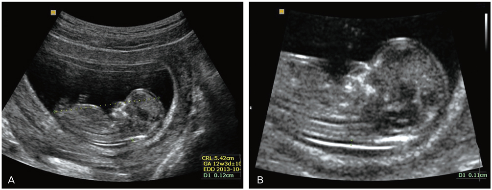
Fig. 1 Measurement of the (A) first measured value (1MV) and (B) second measured value (2MV). (A) The 1MV was measured in the ordinary view for measurement of crown rump length. (B) The 2MV was measured according to the Fetal Medicine Foundation guideline.
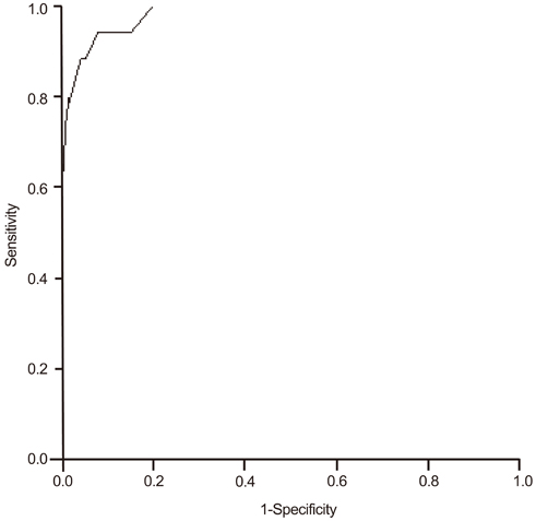
Fig. 2 Receiver operating characteristic curve that describes the performance of the first measured value in the identification of the 95th percentile or more second measured value (area under curve, 0.98; standard error, 0.011; P<0.001).
Reference
-
1. Nicolaides KH, Azar G, Byrne D, Mansur C, Marks K. Fetal nuchal translucency: ultrasound screening for chromosomal defects in first trimester of pregnancy. BMJ. 1992; 304:867–869.2. Pandya PP, Snijders RJ, Johnson SP, De Lourdes Brizot M, Nicolaides KH. Screening for fetal trisomies by maternal age and fetal nuchal translucency thickness at 10 to 14 weeks of gestation. Br J Obstet Gynaecol. 1995; 102:957–962.3. Sebire NJ, Snijders RJ, Hughes K, Sepulveda W, Nicolaides KH. Screening for trisomy 21 in twin pregnancies by maternal age and fetal nuchal translucency thickness at 10-14 weeks of gestation. Br J Obstet Gynaecol. 1996; 103:999–1003.4. Hyett JA, Moscoso G, Nicolaides KH. First-trimester nuchal translucency and cardiac septal defects in fetuses with trisomy 21. Am J Obstet Gynecol. 1995; 172:1411–1413.5. Galindo A, Comas C, Martnez JM, Gutirrez-Larraya F, Carrera JM, Puerto B, et al. Cardiac defects in chromosomally normal fetuses with increased nuchal translucency at 10-14 weeks of gestation. J Matern Fetal Neonatal Med. 2003; 13:163–170.6. Makrydimas G, Sotiriadis A, Huggon IC, Simpson J, Sharland G, Carvalho JS, et al. Nuchal translucency and fetal cardiac defects: a pooled analysis of major fetal echocardiography centers. Am J Obstet Gynecol. 2005; 192:89–95.7. Haak MC, Bartelings MM, Gittenberger-De Groot AC, Van Vugt JM. Cardiac malformations in first-trimester fetuses with increased nuchal translucency: ultrasound diagnosis and postmortem morphology. Ultrasound Obstet Gynecol. 2002; 20:14–21.8. Sebire NJ, Snijders RJ, Davenport M, Greenough A, Nicolaides KH. Fetal nuchal translucency thickness at 10-14 weeks' gestation and congenital diaphragmatic hernia. Obstet Gynecol. 1997; 90:943–946.9. Kagan K, Avgidou K, Molina F, Gajewska K, Nicolaides K. Relation between increased fetal nuchal translucency thickness and chromosomal defects. Obstet Gynecol. 2006; 107:6–10.10. Souka AP, Snijders RJ, Novakov A, Soares W, Nicolaides KH. Defects and syndromes in chromosomally normal fetuses with increased nuchal translucency thickness at 10-14 weeks of gestation. Ultrasound Obstet Gynecol. 1998; 11:391–400.11. Brambati B, Cislaghi C, Tului L, Alberti E, Amidani M, Colombo U, et al. First-trimester Down's syndrome screening using nuchal translucency: a prospective study in patients undergoing chorionic villus sampling. Ultrasound Obstet Gynecol. 1995; 5:9–14.12. Hadlock FP, Shah YP, Kanon DJ, Lindsey JV. Fetal crown-rump length: reevaluation of relation to menstrual age (5-18 weeks) with high-resolution real-time US. Radiology. 1992; 182:501–505.13. Maslovitz S, Yaron Y, Fait G, Gull I, Wolman I, Jaffa A, et al. Feasibility of nuchal translucency in triplet pregnancies. J Ultrasound Med. 2004; 23:501–504.14. Fetal Medicine Foundation. Online education: the 11-13 weeks scan [Internet]. London: Fetal Medicine Foundation;2004. cited 2010 Sep 12. Available from: http://www.fetalmedicine.com.15. Chung JH, Yang JH, Song MJ, Cho JY, Lee YH, Park SY, et al. The distribution of fetal nuchal translucency thickness in normal Korean fetuses. J Korean Med Sci. 2004; 19:32–36.16. Palomaki GE, Lee JE, Canick JA, McDowell GA, Donnenfeld AE. ACMG Laboratory Quality Assurance Committee. Technical standards and guidelines: prenatal screening for Down syndrome that includes first-trimester biochemistry and/or ultrasound measurements. Genet Med. 2009; 11:669–681.17. Teoh M, Meagher SE, Choong S, Shekleton P, Wallace EM. The effect of image size on screen-positive rates for nuchal translucency screening. BJOG. 2006; 113:479–481.18. Hsu JJ, Chiang CH, Hsieh CC, Hsieh TT. The influence of image magnification in first-trimester screening for Down syndrome by fetal nuchal translucency in Asians. Prenat Diagn. 2004; 24:1007–1012.19. Edwards A, Mulvey S, Wallace EM. The effect of image size on nuchal translucency measurement. Prenat Diagn. 2003; 23:284–286.
- Full Text Links
-
- Actions
-
Cited
- CITED
-
- Close
- Share
-
- Similar articles
-
- Changes of Nuchal Translucency in Early Normal Fetuses
- Fetal Nuchal Translucency Measurement for Detection of Chromosomal Abnormalities in the First Trimester of High Risk Pregnancy
- Nuchal Translucency Measurement in Normal Fetuses at 10 - 14 Weeks of Gestation I
- Prenatal chromosomal microarray analysis of fetus with increased nuchal translucency
- Management of pregnancies with increased fetal nuchal translucency in the first trimester
