Radiation-induced eosinophilic, polymorphic, and pruritic eruption in a pig skin model
- Affiliations
-
- 1Laboratory of Radiation Exposure & Therapeutics, Korea Institute of Radiological & Medical Sciences (KIRAMS), Seoul, Korea. sslee@kcch.re.kr, jskim@dirams.re.kr
- 2Research Center, Dongnam Institute of Radiological & Medical Sciences (DIRAMS), Busan, Korea.
- KMID: 2312147
- DOI: http://doi.org/10.5625/lar.2015.31.4.204
Abstract
- Eosinophilic, polymorphic and pruritic eruption associated with radiotherapy (EPPER) can occur in cancer patients after irradiation. In this study, we characterized the clinical and histopathological features of pig skin that developed widespread polymorphic and pruritic skin lesions following localized 50 Gy gamma-irradiation. The pigs developed pruritus 5-7 weeks after irradiation, and infiltration of the dermis by eosinophils was detected 4-7 weeks after irradiation. The irradiated animals also showed transiently increased numbers of peripheral eosinophils 5-7 weeks after treatment. Irradiation induced desquamation after 2-4 weeks, which and the desquamation gradually resolved after 7 weeks. These pathological changes correspond to those seen in irradiated human skin, indicating that this model could be useful for elucidating the pathogenesis of EPPER and for developing therapeutic and prophylactic methods.
Keyword
Figure
-
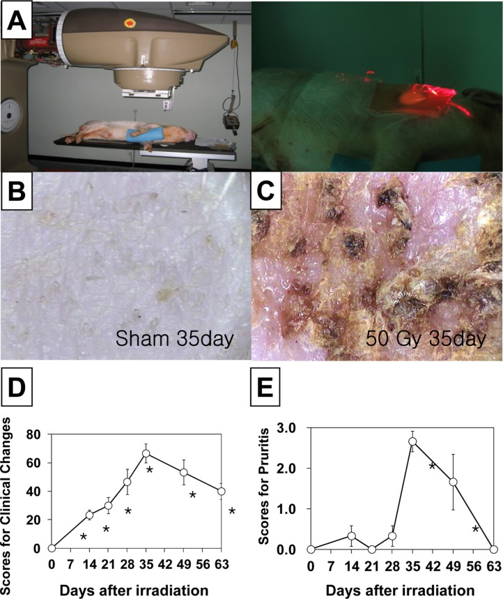
Figure 1 Clinical changes in the skin of a minipig following a 50 Gy irradiation. A. Focal gamma irradiation (50 Gy) was applied to dorsal skin of mini-pigs. B. Sham (0 Gy) control and C. 35 days after irradiation (50 Gy). D. Time-dependent changes in the skin following irradiation. E. Time-dependent development of pruritus following irradiation. The data is reported as the mean±SEM. *P<0.05 vs. sham controls.
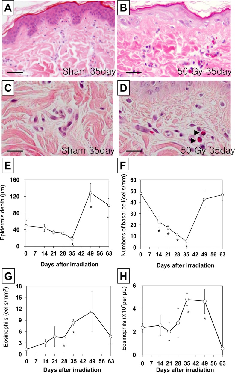
Figure 2 Histological changes in the skin in a hematoxylin and eosin stained section of minipig skin following a 50 Gy irradiation. A and C Skin section from the sham group representative of normal morphology. B and D Skin section from pigs 35 days after irradiation. Basal cell density and epidermal thickness decreased, and eosinophil infiltration increased in the skin 35 days after irradiation. A and B magnification, ×200. Scale bars in A and B: 50 µm. C and D magnification, ×1000. Scale bars in C and D: 20 µm. Time-dependent changes in basal cell density (E), epidermal thickness (F), and the number of dermal (G) and peripheral (H) eosinophils in minipig skin following acute irradiation.. The data is reported as the mean±SEM. *P<0.05 vs. sham controls.
Reference
-
1. Davis J, Pack GT. Erythema multiforme following deep x-ray therapy. AMA Arch Derm Syphilol. 1952; 66(1):41–48. PMID: 14932502.
Article2. Dedick AP Jr, Whelan VM. Generalized skin reaction following deep x-ray therapy. Radiology. 1959; 72(5):751–753. PMID: 13658411.
Article3. Duschet P, Schwarz T, Gschnait F. Bullous pemphigoid after radiation therapy. J Am Acad Dermatol. 1988; 18(2):441–444. PMID: 2963841.
Article4. García-Donoso C, Tardío JC, Arias D, Romero A, Borbujo JM. Eosinophilic, polymorphic and pruritic eruption associated with radiotherapy (EPPER) in two patients with breast tumour. J Eur Acad Dermatol Venereol. 2007; 21(8):1102–1104. PMID: 17714133.
Article5. Lee DJ, Kim YC. A pustular form of eosinophilic, polymorphic, and pruritic eruption associated with radiotherapy. J Am Acad Dermatol. 2011; 65(2):e51–e53. PMID: 21763551.
Article6. Rueda RA, Valencia IC, Covelli C, Escobar C, Alzate A, Saldarriaga B, Sanclemente G, Blank A, Falabella R. Eosinophilic, polymorphic, and pruritic eruption associated with radiotherapy. Arch Dermatol. 1999; 135(7):804–810. PMID: 10411155.
Article7. Lam Cham Kee HX, Charra-Brunaud C, Cuny JF, Reigneau M, Vogin G, Peiffert D. [Case report of EPPER Syndrome (eosinophilic polymorphic pruritic eruption associated with radiotherapy) in a patient treated against endometrial cancer]. Cancer Radiother. 2013; 17(1):54–57. PMID: 23291008.8. Shimamoto N, Shirase T, Yoshikawa Y. Vesicular form of eosinophilic, polymorphic, and pruritic eruption associated with radiotherapy confined to the irradiated area. J Dermatol. 2013; 40(3):228–229. PMID: 23289628.
Article9. Sullivan TP, Eaglstein WH, Davis SC, Mertz P. The pig as a model for human wound healing. Wound Repair Regen. 2001; 9(2):66–76. PMID: 11350644.
Article10. Kim JS, Rhim KJ, Jang WS, Lee SJ, Son Y, Lee SS, Park S, Lim SM. β-irradiation (1Ho patch)-induced skin injury in mini-pigs: effects on NF-κB and COX-2 expression in the skin. J Vet Sci. 2015; 16(1):1–9. PMID: 24962420.11. Hill PB, Lau P, Rybnicek J. Development of an owner-assessed scale to measure the severity of pruritus in dogs. Vet Dermatol. 2007; 18(5):301–308. PMID: 17845617.
Article12. Spry CJ, Davies J, Tai PC, Olsen EG, Oakley CM, Goodwin JF. Clinical features of fifteen patients with the hypereosinophilic syndrome. Q J Med. 1983; 52(205):1–22. PMID: 6878618.
- Full Text Links
-
- Actions
-
Cited
- CITED
-
- Close
- Share
-
- Similar articles
-
- Eosinophilic, Polymorphic, and Pruritic Eruption Associated with Radiotherapy in a Patient with Parotid Gland Cancer
- A Case of Eosinophilic, Polymorphic, and Pruritic Eruption Associated with Radiotherapy in a Patient with Breast Cancer
- Dermatoses of Pregnancy: Clues to Diagnosis, Fetal Risk and Therapy
- Two Cases of Pruritic Urticarial Papules and Plaques of Pregnancy in the Postpartum
- A Case of Polymorphic Pemphigoid
