Lab Anim Res.
2013 Sep;29(3):174-177. 10.5625/lar.2013.29.3.174.
Development of osteoporosis animal model using micropigs
- Affiliations
-
- 1Laboratory Animal Center, Korea Research Institute of Bioscience and Biotechnology (KRIBB), Daejeon, Korea.
- 2Medikinetics Co., Ltd., Pyeongtaek, Korea. osm280@daum.net bhhyun@kbio.kr
- 3Laboratory Animal Center, Osong Medical Innovation Foundation (KBIO), Chungbuk, Korea. osm280@daum.net bhhyun@kbio.kr
- KMID: 2312111
- DOI: http://doi.org/10.5625/lar.2013.29.3.174
Abstract
- Osteoporosis is a known major health problem and a serious disease of the bone, there has been a great need to develop more and newer animal models for this disease. Among animal models used for testing drug efficacy, the minipig model has become useful and effective due to its close similarity with humans (validity), particularly with the pharmacokinetics of compounds via subcutaneous administration, the structure and function of the organs, the morphology of bone and the overall metabolic nature. Based on these advantages, we sought to develop a new animal model of osteoporosis using micropig, which differs from other miniature pigs in the genetic background. Female micropigs were used for the induction of a moderate osteoporosis model by bilateral ovariectomy (OVX) and compared with shamoperated animals. For osteoporosis evaluation, clinical biomarkers such as blood osteocalcin (OSC) and parathyroid hormone (PTH) levels were measured, as well as bone mineral density (BMD) using micro-computed tomography (micro-CT). Compared to sham, OVX animals have decreased blood OSC level, while the blood PTH level increased in blood sera. In addition, we observed the significantly decreased BMDs of tibia region in OVX animals. Based on these results, we report that the micropig model developed in this study can be used to develop a new and effective medical method for diagnosis and treatment of osteoporosis.
Keyword
MeSH Terms
Figure
-
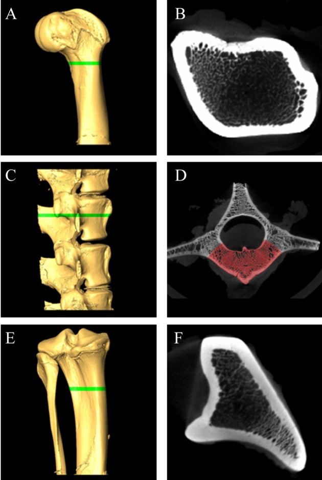
Figure 1 Micro-CT analysis of the femur, lumbar, and tibia bone of micorpig. 3D volume rendering images (A,C,E) and corresponding cross section images (B,D,F). (A) and (B), femur, (C) and (D), lumbar, (E) and (F), tibia. Green bars of A, C, E are designated ROIs. (E) Ulnar portion was excluded for ROI. (D) In lumbar's cross section, only red area (vertebral body portion) was calculated for BMD because other areas are included in transverse process and spinal process. Femur (B) and tibia (F) are calculated whole cross section. ROI; region of interest, BMD; bone mineral density.
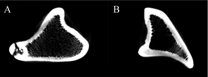
Figure 2 Corresponding cross section images of the tibia bone in sham operated (A) and ovariectomized (B) groups.
Reference
-
1. Miller ER, Ullrey DE. The pig as a model for human nutrition. Annu Rev Nutr. 1987; 7:361–382. PMID: 3300739.
Article2. Hönig JF, Merten HA. Subperiosteal versus epiperiosteal forehead augmentation with hydroxylapatite for aesthetic facial contouring: experimental animal investigation and clinical application. Aesthetic Plast Surg. 1993; 17(2):93–98. PMID: 8390778.
Article3. Swindle MM, Smith AC. Comparative anatomy and physiology of the pig. Scand J Lab Anim Sci. 1998; 25:11–21.4. Scholz-Ahrens KE, Delling G, Stampa B, Helfenstein A, Hahne HJ, Açil Y, Timm W, Barkmann R, Hassenpflug J, Schrezenmeir J, Glüer CC. Glucocorticosteroid-induced osteoporosis in adult primiparous Göttingen miniature pigs: effects on bone mineral and mineral metabolism. Am J Physiol Endocrinol Metab. 2007; 293(1):E385–E395. PMID: 17456640.
Article5. Akahoshi S, Sakai A, Arita S, Ikeda S, Morishita Y, Tsutsumi H, Ito M, Shiraishi A, Nakamura T. Modulation of bone turnover by alfacalcidol and/or alendronate does not prevent glucocorticoid-induced osteoporosis in growing minipigs. J Bone Miner Metab. 2005; 23(5):341–350. PMID: 16133683.
Article6. Jee WS, Yao W. Overview: animal models of osteopenia and osteoporosis. J Musculoskelet Neuronal Interact. 2001; 1(3):193–207. PMID: 15758493.7. Turner AS. Animal models of osteoporosis--necessity and limitations. Eur Cell Mater. 2001; 1:66–81. PMID: 14562261.8. Yoon KH, Cho DC, Yu SH, Kim KT, Jeon Y, Sung JK. The Change of Bone Metabolism in Ovariectomized Rats : Analyses of MicroCT Scan and Biochemical Markers of Bone Turnover. J Korean Neurosurg Soc. 2012; 51(6):323–327. PMID: 22949959.
Article9. Jochems C, Islander U, Erlandsson M, Verdrengh M, Ohlsson C, Carlsten H. Osteoporosis in experimental postmenopausal polyarthritis: the relative contributions of estrogen deficiency and inflammation. Arthritis Res Ther. 2005; 7(4):R837–R843. PMID: 15987485.10. Mosekilde L, Weisbrode SE, Safron JA, Stills HF, Jankowsky ML, Ebert DC, Danielsen CC, Sogaard CH, Franks AF, Stevens ML, Paddock CL, Boyce RW. Calcium-restricted ovariectomized Sinclair S-1 minipigs: an animal model of osteopenia and trabecular plate perforation. Bone. 1993; 14(3):379–382. PMID: 8363881.
Article11. Boyce RW, Ebert DC, Youngs TA, Paddock CL, Mosekilde L, Stevens ML, Gundersen HJ. Unbiased estimation of vertebral trabecular connectivity in calcium-restricted ovariectomized minipigs. Bone. 1995; 16(6):637–642. PMID: 7669440.
Article12. Tsutsumi H, Katagiri K, Takeda S, Nasu T, Igarashi S, Tanigawa M, Mamba K. Standardized data and relationship between bone growth and bone metabolism in female Göttingen minipigs. Exp Anim. 2004; 53(4):331–337. PMID: 15297706.13. Farrugia W, Fortune CL, Heath J, Caple IW, Wark JD. Osteocalcin as an index of osteoblast function during and after ovine pregnancy. Endocrinology. 1989; 125(3):1705–1710. PMID: 2788077.
Article14. Genant HK, Jiang Y. Advanced imaging assessment of bone quality. Ann N Y Acad Sci. 2006; 1068:410–428. PMID: 16831940.
Article15. Ikeda S, Morishita Y, Tsutsumi H, Ito M, Shiraishi A, Arita S, Akahoshi S, Narusawa K, Nakamura T. Reductions in bone turnover, mineral, and structure associated with mechanical properties of lumbar vertebra and femur in glucocorticoid-treated growing minipigs. Bone. 2003; 33(5):779–787. PMID: 14623053.
Article
- Full Text Links
-
- Actions
-
Cited
- CITED
-
- Close
- Share
-
- Similar articles
-
- Evaluation of the lateral ventricle using MRI in normal micropigs
- The Effects of Combination Therapy of Cathepsin K Inhibitor and PTH on Change of Bone Mineral Density in Animal Model of Osteoporosis
- Comparison of cardiac function and coronary angiography between conventional pigs and micropigs as measured by multidetector row computed tomography
- Cerebellar maturation ratio of forebrain and brainstem at magnetic resonance imaging in the micropig
- Letter: The Effects of Combination Therapy of Cathepsin K Inhibitor and PTH on Change in Bone Mineral Density in an Animal Model of Osteoporosis
