Evaluation of Renal Microvasculature Using Micro-computed Tomography in Rat
- Affiliations
-
- 1Department of Urology, Wonkwang University School of Medicine, Iksan, Korea. seraph@wonkwang.ac.kr
- 2Department of Radiology, Wonkwang University School of Medicine and the Institute for Radiological Imaging Science, Iksan, Korea.
Abstract
-
PURPOSE: Rodent models that mimic human renal diseases are being increasingly recognized as powerful tools in the development of new drugs and for evaluating the efficacy of novel therapeutics in a preclinical setting. However, there are few reports on microvasculature imaging of the urinary system in small animals. An experimental study was performed to evaluate the microvasculature in a rat kidney using micro- computed tomography(CT) with three-dimensional images.
MATERIALS AND METHODS
Five Sprague-Dawley male rats(age: 10-12 weeks, weight: 200-250g) underwent a laparotomy under anesthesia with an intramuscular injection of 0.5cc xylazine hydrochloride and ketamine mixed solution(1:10). After ligation of the abdominal aorta and inferior vena cava immediately above the renal artery, a 24 gazed catheter was inserted into the abdominal aorta. A physiological solution and heparin(500U) were infused through the catheter to flush the blood from the renal vasculature. The kidney was enhanced using self-made contrast material. The excised kidney was frozen for the micro-CT scan.
RESULTS
The mean longitudinal diameter and weight of the 10 resected kidneys was 1.95+/-0.15cm and 2.0+/-0.28g, respectively. The images were represented by three-dimensional arrays of cubic voxels with opacities in the blood vessels. In the section taken from the arrays, four regions of the kidney could be identified easily by their characteristic vascular features.
CONCLUSIONS
Micro-CT is a promising method for evaluating the renal microvascular architecture in a rat kidney. It can for the foundation of an experimental study aimed at providing quantitative information on the urinary system in a rodent model.
MeSH Terms
Figure
-
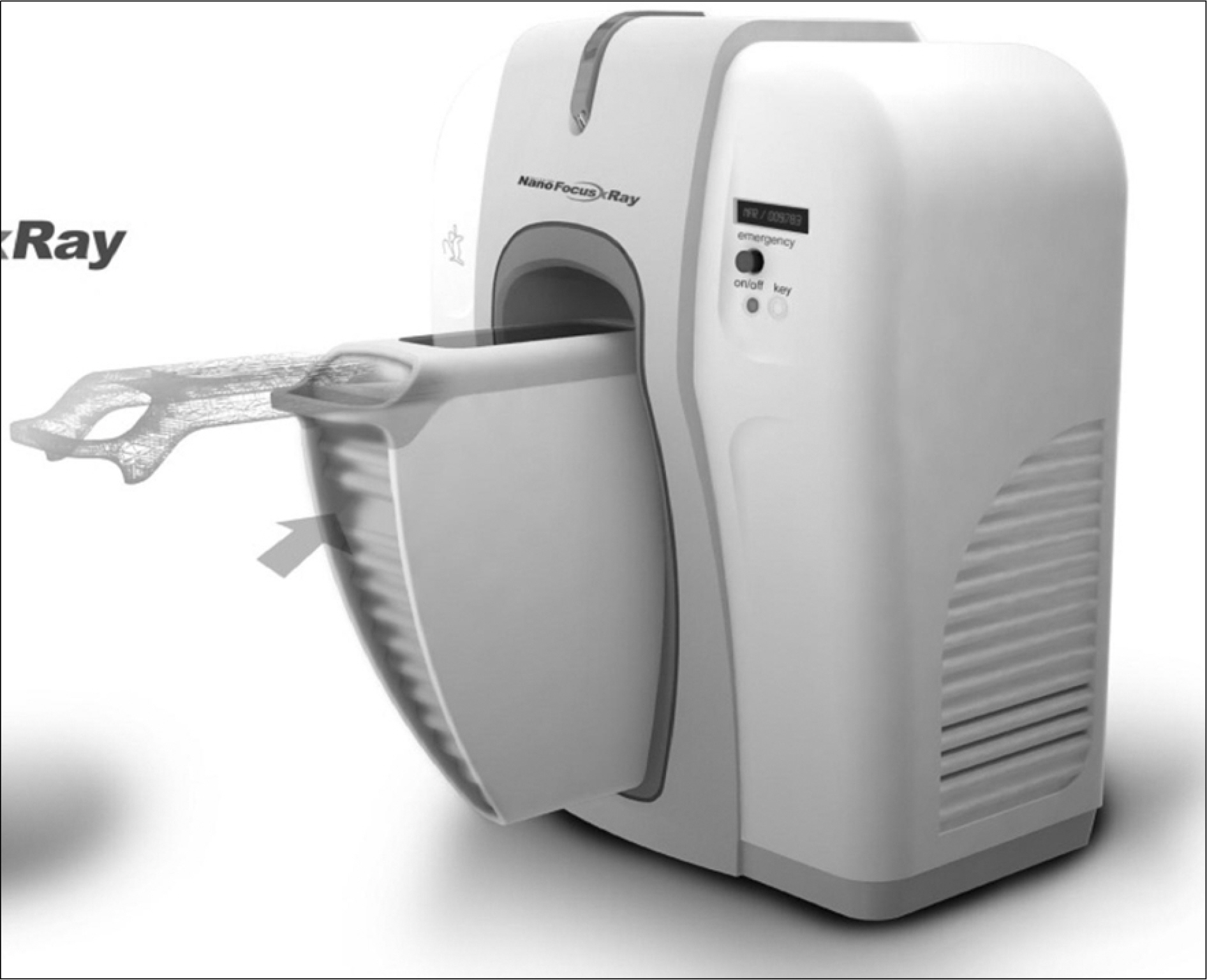
Fig. 1. Photograph of the developed micro-computed tomography system for in vivo small animal imaging.

Fig. 2. The preparation steps for micro-computed tomography (CT) scanning. (A) Laparotomy was performed after anesthesia by an intramuscular injection of 0.5cc xylazine hydrochloride and ketamine mixed solution (1:10). (B) After the ligation of the abdominal aorta and inferior vena cava immediately above the renal artery, a 24 gauged catheter was inserted into the abdominal aorta at 1cm below to the renal artery. (C) A physiologic solution (10cc) and heparin (500U) were infused through the catheter to flush the blood from the renal vasculature. (D) The colors of the kidneys were changed because of the fixation with the contrast material, gelatin and barium sulfate (BaSO4). (E) The excised kidney was frozen for the micro-CT scan.
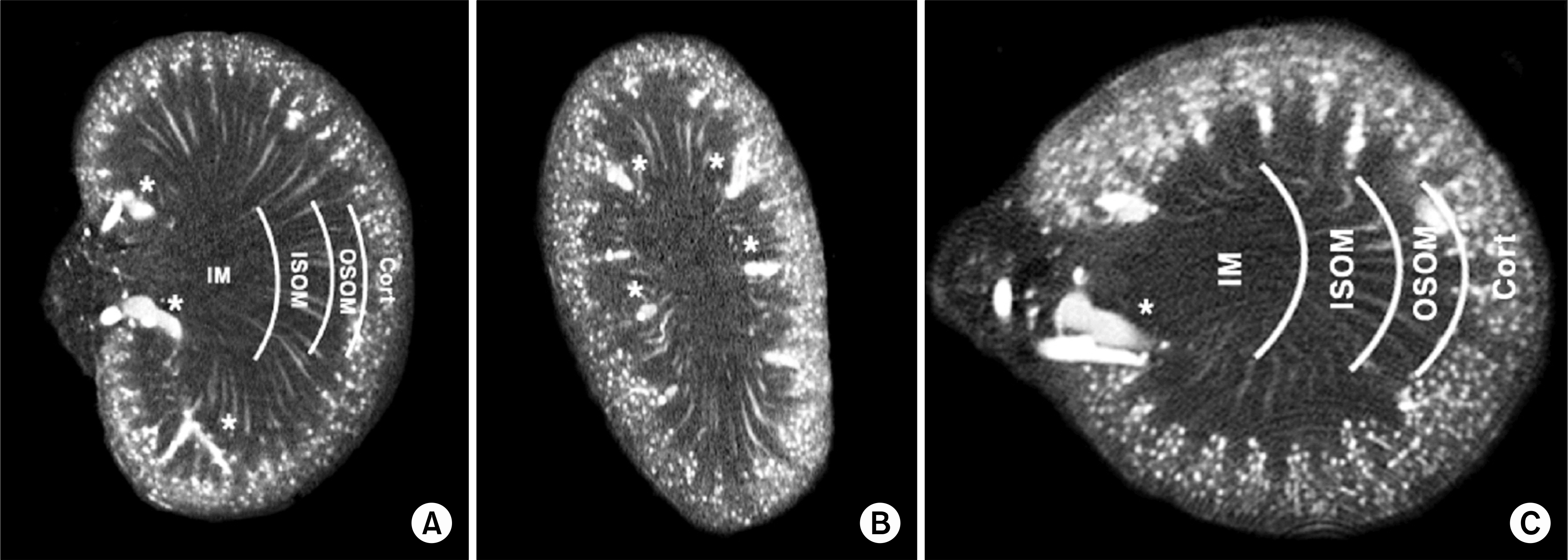
Fig. 3. Coronal (A), sagittal (B), and transverse (C) sections through a volume image of a rat kidney. The cortex (Cort), outer stripe of the outer medulla (OSOM), inner stripe of outer medulla (ISOM), and the inner medulla (IM) were distinguished by their vascular features. Interlobar vessels (*) are evident in the sections.
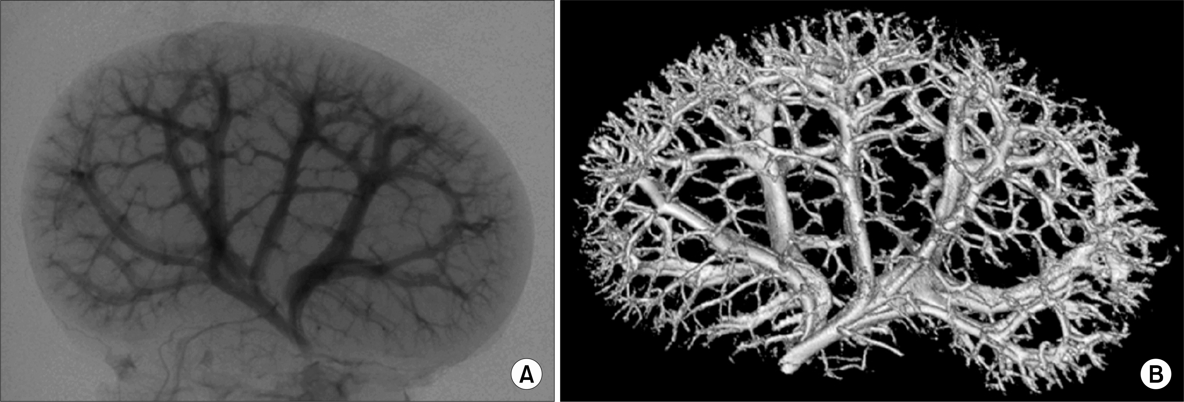
Fig. 4. Renal microvasculature images of the ex-vivo mouse in the micro-computed tomography (CT). (A) projection image, (B) 3 dimensional-CT reconstruction image.
Reference
-
1.Seo IY., Jeong CS., Rim JS. The anti-inflammatory effect of cobalt-protoporphyrin for rats with epididymitis induced by Escherichia coli infection. Korean J Urol. 2006. 47:656–60.2.Nam JK., Lee SD., Kim BO. HIF-1alpha, iNOS and VEGF expression of contralateral kidney during acute stage in complete ureteral obstruction of rat model. Korean J Urol. 2005. 46:518–25.3.Ritman EL. Micro-computed tomography-current status and developments. Annu Rev Biomed Eng. 2004. 6:185–208.
Article4.Feldkamp LA., Goldstein SA., Parfitt AM., Jesion G., Kleerekoper M. The direct examination of three-dimensional bone architecture in vitro by computed tomography. J Bone Miner Res. 1989. 4:3–11.
Article5.Paulus MJ., Gleason SS., Kennel SJ., Hunsicker PR., Johnson DK. High resolution X-ray computed tomography: an emerging tool for small animal cancer research. Neoplasia. 2000. 2:62–70.
Article6.Fortepiani LA., Ruiz MC., Passardi F., Bentley MD., Garcia-Estan J., Ritman EL, et al. Effect of losartan on renal microvasculature during chronic inhibition of nitric oxide visualized by micro-CT. Am J Physiol Renal Physiol. 2003. 285:F852–60.
Article7.Feldkamp LA., Davis LC., Kress JW. Practical cone-beam algorithm. J Opt Soc Am A. 1984. 1:612–9.
Article8.Toyota E., Ogasawara Y., Fujimoto K., Kajita T., Shigeto F., Asano T, et al. Global heterogeneity of glomerular volume distribution in early diabetic nephropathy. Kidney Int. 2004. 66:855–61.
Article9.Gossl M., Bentley MD., Lerman LO. Review-3D micro CT imaging of renal micro-structural changes. Nephron Clin Pract. 2006. 103:c66–70.10.Ludders JW., Wilson JW., Ribble GA. Microangiography and correlated histology: a research technique for examining renal microcirculation. Am J Vet Res. 1985. 46:2536–8.11.Evan AP., Dail WG Jr. Efferent arterioles in the cortex of the rat kidney. Anat Rec. 1977. 187:135–45.
Article12.Sung KT., Yoon JB. Comparative studies of the renal vasculature of the human and the experimental animals by renal microangiography and corrosion casts. Korean J Urol. 1990. 31:471–80.13.Bentley MD., Ortiz MC., Ritman EL., Romero JC. The use of microcomputed tomography to study microvasculature in small rodents. Am J Physiol Regul Integr Comp Physiol. 2002. 282:F1267–79.
Article14.Garcia-Sanz A., Rodriguez-Barbero A., Bentley MD., Ritman EL., Romero JC. Three-dimensional microcomputed tomography of renal vasculature in rats. Hypertension. 1998. 31:440–4.
Article15.Ritman EL., Jorgensen SM., Lund PE., Thomas PJ., Dunsmuir JH., Romero JC, et al. Synchrotron-based micro-CT of in-situ biological basic functional units and their integration. Proc Soc Photo Opt Instrum Eng. 1997. 3149:13–24.16.Lemley KV., Kriz W. Structure and function of the renal vasculature. Tisher CC, Brenner BM, editors. editors.Renal pathology with clinical and functional correlations. 1st ed.Philadelphia: Lippincott Williams & Wilkins;1989. 926-64.17.Pfaller W. Structure function correlation on rat kidney. Quantitative correlation of structure and function in the normal and injured rat kidney. Adv Anat Embryol Cell Biol. 1982. 70:1–106.18.Jorgensen SM., Demirkaya O., Ritman EL. Three-dimensional imaging of vasculature and parenchyma in intact rodent organs with X-ray micro-CT. Am J Physiol. 1998. 275:H1103–14.
- Full Text Links
-
- Actions
-
Cited
- CITED
-
- Close
- Share
-
- Similar articles
-
- Micro-computed tomography analysis of changes in the periodontal ligament and alveolar bone proper induced by occlusal hypofunction of rat molars
- Evaluation of 3 Cases of Renal Injuries with Dynamic Computed Tomography
- The evaluation of the correlation between histomorphometric analysis and micro-computed tomography analysis in AdBMP-2 induced bone regeneration in rat calvarial defects
- Recovery of Renal Microvasculature and Expression of VEGF in Experimental Thrombotic Microangiopathy
- Recent advances in airway imaging using micro-computed tomography and computed tomography for chronic obstructive pulmonary disease
