Micro-computed tomography analysis of changes in the periodontal ligament and alveolar bone proper induced by occlusal hypofunction of rat molars
- Affiliations
-
- 1Orthodontic Science, Department of Orofacial Development and Function, Division of Oral Health Sciences, Graduate School, Tokyo Medical and Dental University, Tokyo, Japan. y.shimizu.orts@tmd.ac.jp
- KMID: 1726420
- DOI: http://doi.org/10.4041/kjod.2014.44.5.263
Abstract
OBJECTIVE
To three-dimensionally elucidate the effects of occlusal hypofunction on the periodontal ligament and alveolar bone proper of rat molars by micro-computed tomography (micro-CT).
METHODS
Occlusal function in the molar area was restricted by attaching an anterior bite plate on the maxillary incisors and a metal cap on the mandibular incisors of 5-week-old male Wistar rats for 1 week. The periodontal ligament space and alveolar bone proper around roots of the mandibular first molar were assessed by histology and micro-CT.
RESULTS
The periodontal ligament space was narrower and the alveolar bone proper was sparser and less continuous in the hypofunction group than in the control group. Further, both the volume of the periodontal ligament and the volumetric ratio of the alveolar bone proper to the total tissue in the region of interest were significantly lower in the hypofunction group (p < 0.05).
CONCLUSIONS
Occlusal hypofunction induces atrophic changes in the periodontal ligament and alveolar bone proper of rat molars.
Keyword
Figure
-

Figure 1 A, The experimental model; B, histological observational area (rectangular area); C, representative hematoxylin and eosin-stained sections (Scale = 250 µm). M, Mesial; D, distal; PL, periodontal ligament; AP, alveolar bone proper; AB, inter-radicular alveolar bone.
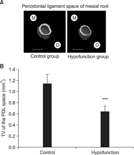
Figure 2 A, Three-dimensional reconstructed images of the periodontal ligament (PDL) space (Scale = 1 mm); B, comparison of tissue volume (TV) around the mesial root of the mandibular first molar. ***p < 0.001 by the Mann-Whitney U-test. M, Mesial; D, distal.

Figure 3 A, Three-dimensional reconstructed images of the periodontal ligament (PDL) space (Scale = 1 mm); B, comparison of tissue volume (TV) around the distal root of the mandibular first molar. ***p < 0.001 by the Mann-Whitney U-test. M, Mesial; D, distal.

Figure 4 A, Three-dimensional reconstructed images of the alveolar bone proper (Scale = 1 mm); B, comparison of the bone-to-tissue volume (BV/TV) ratio around the mesial root of the mandibular first molar. *p < 0.05 by the Mann-Whitney U-test. M, Mesial; D, distal.
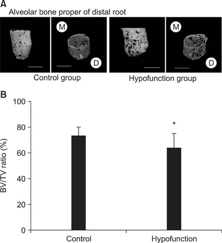
Figure 5 A, Three-dimensional reconstructed images of the alveolar bone proper (Scale = 1 mm); B, comparison of the bone-to-tissue volume (BV/TV) ratio around the distal root of the mandibular first molar. *p < 0.05 by the Mann-Whitney U-test. M, Mesial; D, distal.
Cited by 2 articles
-
Three-dimensional structural analysis of the morphological condition of the alveolar bone before and after orthodontic treatment
Yasuhiro Shimizu, Takashi Ono
Korean J Orthod. 2017;47(6):394-400. doi: 10.4041/kjod.2017.47.6.394.Long-term survival of retained deciduous mandibular second molars and maxillary canine incorporated into final occlusion
Soonshin Hwang, Yoon Jeong Choi, Chooryung J. Chung, Kyung-Ho Kim
Korean J Orthod. 2017;47(5):323-333. doi: 10.4041/kjod.2017.47.5.323.
Reference
-
1. Gault P, Black A, Romette JL, Fuente F, Schroeder K, Thillou F, et al. Tissue-engineered ligament: implant constructs for tooth replacement. J Clin Periodontol. 2010; 37:750–758.
Article2. Sebaoun JD, Kantarci A, Turner JW, Carvalho RS, Van Dyke TE, Ferguson DJ. Modeling of trabecular bone and lamina dura following selective alveolar decortication in rats. J Periodontol. 2008; 79:1679–1688.
Article3. Devlin H, Sloan P. Early bone healing events in the human extraction socket. Int J Oral Maxillofac Surg. 2002; 31:641–645.
Article4. Gay IC, Chen S, MacDougall M. Isolation and characterization of multipotent human periodontal ligament stem cells. Orthod Craniofac Res. 2007; 10:149–160.
Article5. Hayashi Y, Iida J, Warita H, Soma K. Effects of occlusal hypofunction on the microvasculature and endothelin expression in the periodontal ligaments of rat molars. Orthod Waves. 2001; 60:373–380.6. Tanaka A, Iida J, Soma K. Effect of hypofunction on the microvasculature in the periodontal ligament of the rat molar. Orthodontic Waves. 1998; 57:180–188.7. Muramoto T, Takano Y, Soma K. Time-related changes in periodontal mechanoreceptors in rat molars after the loss of occlusal stimuli. Arch Histol Cytol. 2000; 63:369–380.
Article8. Enokida M, Kaneko S, Yanagishita M, Soma K. Inference of occlusal stimuli on the remodelling of alveolar bone in a rat hypofunction-recovery model. J Oral Biosci. 2005; 47:321–334.
Article9. Shimomoto Y, Chung CJ, Iwasaki-Hayashi Y, Muramoto T, Soma K. Effects of occlusal stimuli on alveolar/jaw bone formation. J Dent Res. 2007; 86:47–51.
Article10. Shimizu Y, Hosomichi J, Kaneko S, Shibutani N, Ono T. Effect of sympathetic nervous activity on alveolar bone loss induced by occlusal hypofunction in rats. Arch Oral Biol. 2011; 56:1404–1411.
Article11. Motokawa M, Terao A, Karadeniz EI, Kaku M, Kawata T, Matsuda Y, et al. Effects of long-term occlusal hypofunction and its recovery on the morphogenesis of molar roots and the periodontium in rats. Angle Orthod. 2013; 83:597–604.
Article12. Watarai H, Warita H, Soma K. Effect of nitric oxide on the recovery of the hypofunctional periodontal ligament. J Dent Res. 2004; 83:338–342.
Article13. Bourrin S, Palle S, Genty C, Alexandre C. Physical exercise during remobilization restores a normal bone trabecular network after tail suspension-induced osteopenia in young rats. J Bone Miner Res. 1995; 10:820–828.
Article14. Vignery A, Baron R. Dynamic histomorphometry of alveolar bone remodeling in the adult rat. Anat Rec. 1980; 196:191–200.
Article15. Johnson RB. Effect of altered occlusal function on transseptal ligament and new bone thicknesses in the periodontium of the rat. Am J Anat. 1990; 187:91–97.
Article16. Meeran NA. Cellular response within the periodontal ligament on application of orthodontic forces. J Indian Soc Periodontol. 2013; 17:16–20.
Article17. Mayahara K, Yamaguchi A, Takenouchi H, Kariya T, Taguchi H, Shimizu N. Osteoblasts stimulate osteoclastogenesis via RANKL expression more strongly than periodontal ligament cells do in response to PGE(2). Arch Oral Biol. 2012; 57:1377–1384.
Article18. Krishnan V, Davidovitch Z. Cellular, molecular, and tissue-level reactions to orthodontic force. Am J Orthod Dentofacial Orthop. 2006; 129:469.e1–469.e32.
Article
- Full Text Links
-
- Actions
-
Cited
- CITED
-
- Close
- Share
-
- Similar articles
-
- Micro-CT analysis of LPS-induced Alveolar Bone Loss in Diabetic Mice
- Quantification of Microstructures in Mice Alveolar Bone using Micro-computed tomography (microCT)
- Three-dimentional finite element analysis of a mandibular premolar with reduced periodontal support under a non-axial load
- The influence of root surface distance to alveolar bone and periodontal ligament on periodontal wound healing
- The study on the periodontal vascular changes of rat incisors following experimental tooth movement
