Ann Rehabil Med.
2016 Apr;40(2):190-196. 10.5535/arm.2016.40.2.190.
Assessment of Oropharyngeal Dysphagia in Patients With Parkinson Disease: Use of Ultrasonography
- Affiliations
-
- 1Department of Physical Medicine and Rehabilitation, Veterans Health Service Medical Center, Seoul, Korea. khojing@hanmail.net
- KMID: 2309916
- DOI: http://doi.org/10.5535/arm.2016.40.2.190
Abstract
OBJECTIVE
To compare tongue thickness, the shortest hyoid-thyroid approximation (distance between the hyoid bone and thyroid cartilage), and the time interval between the initiation of tongue movement and the time of the shortest hyoid-thyroid approximation, by using ultrasonography in healthy controls and patients with Parkinson disease (PD).
METHODS
Healthy controls and PD patients with dysphagia were compared. Ultrasonography was performed 3 times for the evaluation of tongue thickness, the shortest hyoid-thyroid approximation, and the time between the initiation of tongue movement and the shortest hyoid-thyroid approximation.
RESULTS
A total of 24 healthy controls and 24 PD patients with dysphagia were enrolled. No significant differences were demonstrated between the two groups for the shortest hyoid-thyroid approximation (controls, 1.19±0.34 cm; PD patients, 1.37±0.5 cm; p=0.15) and tongue thickness (controls, 4.42±0.46 cm; PD patients, 4.27±0.51 cm; p=0.3). In contrast, the time to the shortest hyoid-thyroid approximation was significantly different between the two groups (controls, 1.53±0.87 ms; PD patients, 2.4±1.4 ms, p=0.048).
CONCLUSION
Ultrasonography can be useful in evaluating dysphagia in patients with PD by direct visualization and measurement of the hyoid bone. Moreover, ultrasonography might contribute to a greater understanding of the pathophysiology of dysphagia in PD.
Keyword
MeSH Terms
Figure
-
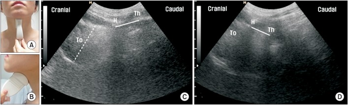
Fig. 1 Longitudinal ultrasonographic images of hyoid-thyroid approximation at rest and during the swallowing state. (A) Longitudinal approach of ultrasonographic probe, anterior view. (B) Longitudinal approach of ultrasonographic probe, lateral view. (C) At rest, the thickest area of the tongue ('To') is shown by the dotted line, and the hyoid-thyroid approximation, which is the distance between the hyoid bone ('H') and the thyroid cartilage ('Th'), is shown by the solid line. (D) During swallowing, hyoid-thyroid approximation occurs with laryngeal elevation. The shortest distance of hyoid-thyroid approximation is shown by the solid line.
Reference
-
1. Jankovic J. Parkinson's disease: clinical features and diagnosis. J Neurol Neurosurg Psychiatry. 2008; 79:368–376. PMID: 18344392.
Article2. Storch A, Schneider CB, Wolz M, Sturwald Y, Nebe A, Odin P, et al. Nonmotor fluctuations in Parkinson disease: severity and correlation with motor complications. Neurology. 2013; 80:800–809. PMID: 23365054.
Article3. Nicaretta DH, Rosso AL, Mattos JP, Maliska C, Costa MM. Dysphagia and sialorrhea: the relationship to Parkinson's disease. Arq Gastroenterol. 2013; 50:42–49. PMID: 23657306.4. Kalf JG, de Swart BJ, Bloem BR, Munneke M. Prevalence of oropharyngeal dysphagia in Parkinson's disease: a meta-analysis. Parkinsonism Relat Disord. 2012; 18:311–315. PMID: 22137459.
Article5. Cereda E, Cilia R, Klersy C, Canesi M, Zecchinelli AL, Mariani CB, et al. Swallowing disturbances in Parkinson's disease: a multivariate analysis of contributing factors. Parkinsonism Relat Disord. 2014; 20:1382–1387. PMID: 25456827.
Article6. Baijens LW, Speyer R, Passos VL, Pilz W, Roodenburg N, Clave P. Swallowing in Parkinson patients versus healthy controls: reliability of measurements in videofluoroscopy. Gastroenterol Res Pract. 2011; 2011:380682. PMID: 21977026.
Article7. Monte FS, da Silva-Junior FP, Braga-Neto P, Nobre e Souza MA, de Bruin VM. Swallowing abnormalities and dyskinesia in Parkinson's disease. Mov Disord. 2005; 20:457–462. PMID: 15625689.
Article8. Troche MS, Okun MS, Rosenbek JC, Musson N, Fernandez HH, Rodriguez R, et al. Aspiration and swallowing in Parkinson disease and rehabilitation with EMST: a randomized trial. Neurology. 2010; 75:1912–1919. PMID: 21098406.
Article9. Logemann JA. Role of the modified barium swallow in management of patients with dysphagia. Otolaryngol Head Neck Surg. 1997; 116:335–338. PMID: 9121786.
Article10. Kuhl V, Eicke BM, Dieterich M, Urban PP. Sonographic analysis of laryngeal elevation during swallowing. J Neurol. 2003; 250:333–337. PMID: 12638025.
Article11. Macrae PR, Doeltgen SH, Jones RD, Huckabee ML. Intra- and inter-rater reliability for analysis of hyoid displacement measured with sonography. J Clin Ultrasound. 2012; 40:74–78. PMID: 21953135.
Article12. Kim JH, Kim MS. Lateral pharyngeal wall motion analysis using ultrasonography in stroke patients with dysphagia. Ultrasound Med Biol. 2012; 38:2058–2064. PMID: 23062372.
Article13. Yabunaka K, Sanada H, Sanada S, Konishi H, Hashimoto T, Yatake H, et al. Sonographic assessment of hyoid bone movement during swallowing: a study of normal adults with advancing age. Radiol Phys Technol. 2011; 4:73–77. PMID: 20945118.
Article14. Huang YL, Hsieh SF, Chang YC, Chen HC, Wang TG. Ultrasonographic evaluation of hyoid-larynx approximation in dysphagic stroke patients. Ultrasound Med Biol. 2009; 35:1103–1108. PMID: 19427098.
Article15. Hsiao MY, Chang YC, Chen WS, Chang HY, Wang TG. Application of ultrasonography in assessing oropharyngeal dysphagia in stroke patients. Ultrasound Med Biol. 2012; 38:1522–1528. PMID: 22698507.
Article16. Volonte MA, Porta M, Comi G. Clinical assessment of dysphagia in early phases of Parkinson's disease. Neurol Sci. 2002; 23(Suppl 2):S121–S122. PMID: 12548373.17. Goetz CG, Poewe W, Rascol O, Sampaio C, Stebbins GT, Counsell C, et al. Movement Disorder Society Task Force report on the Hoehn and Yahr staging scale: status and recommendations. Mov Disord. 2004; 19:1020–1028. PMID: 15372591.18. Morgan A, Ward E, Murdoch B, Bilbie K. Acute characteristics of pediatric dysphagia subsequent to traumatic brain injury: videofluoroscopic assessment. J Head Trauma Rehabil. 2002; 17:220–241. PMID: 12086576.19. Nagaya M, Kachi T, Yamada T, Igata A. Videofluorographic study of swallowing in Parkinson's disease. Dysphagia. 1998; 13:95–100. PMID: 9513304.
Article20. Shawker TH, Sonies B, Hall TE, Baum BF. Ultrasound analysis of tongue, hyoid, and larynx activity during swallowing. Invest Radiol. 1984; 19:82–86. PMID: 6398320.
Article21. Fanucci A, Cerro P, Ietto F, Brancaleone C, Berardi F. Physiology of oral swallowing studied by ultrasonography. Dentomaxillofac Radiol. 1994; 23:221–225. PMID: 7835528.
Article22. Mu L, Sobotka S, Chen J, Su H, Sanders I, Adler CH, et al. Altered pharyngeal muscles in Parkinson disease. J Neuropathol Exp Neurol. 2012; 71:520–530. PMID: 22588389.
Article
- Full Text Links
-
- Actions
-
Cited
- CITED
-
- Close
- Share
-
- Similar articles
-
- Dysphagia in the patients with Parkinson's Disease
- Oropharyngeal Cancer and Dysphagia
- Oro-Pharyngeal Dysphagia in Parkinson's Disease and Related Movement Disorders
- Swallowing Training for Patients with Impaired Cardiopulmonary Function
- Clinical Characteristics and Evaluation of Dysphagia in Patients with Parkinson’s Disease
