Evaluation of canalis basilaris medianus using cone-beam computed tomography
- Affiliations
-
- 1Department of Oral and Maxillofacial Medicine and Diagnostic Sciences, CWRU School of Dental Medicine, Cleveland, OH, USA. azs16@case.edu
- 2Department of Oral Diagnostic Sciences, VCU School of Dentistry, Richmond, VA, USA.
- 3Division of Radiology, University of Pennsylvania School of Dental Medicine, Philadelphia, PA, USA.
- KMID: 2308878
- DOI: http://doi.org/10.5624/isd.2016.46.2.141
Abstract
- The aim of this report is to present two cases of canalis basilaris medianus as identified on cone-beam computed tomography (CBCT) in the base of the skull. The CBCT data sets were sent for radiographic consultation. In both cases, multi-planar views revealed an osseous defect in the base of the skull in the clivus region, the sagittal view showed a unilateral, well-defined, non-corticated, track-like low-attenuation osseous defect in the clivus. The appearance of the defect was highly reminiscent of a fracture of the clivus. The borders of osseous defect were smooth, and no other radiographic signs suggestive of osteolytic destructive processes were noted. Based on the overall radiographic examination, a radiographic impression of canalis basilaris medianus was made. Canalis basilaris medianus is a rare anatomical variant and is generally observed on the clivus. Due to its potential association with meningitis, it should be recognized and reported to avoid potential complications.
Figure
-
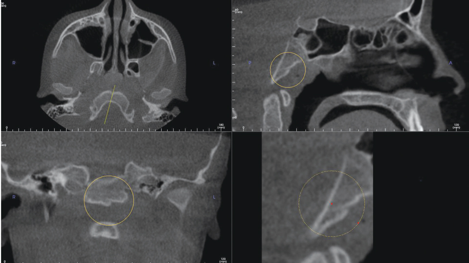
Fig. 1 Multiplanar reconstruction images demonstrate a defect on the basiocciput of the clivus consistent with canalis basilaris medianus in an 11-year-old female.

Fig. 2 A sagittal image demonstrates canalis basilaris medianus of the complete type on the superior aspect of the clivus.
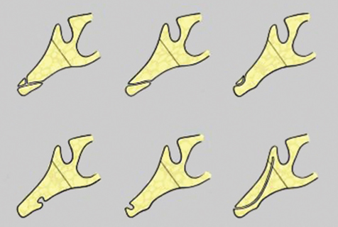
Fig. 3 A schematic diagram depicting the different types of canalis basilaris medianus (CBM). The top row illustrates the complete forms of CBM and the bottom row illustrates incomplete forms of CBM. Top row: bifurcating type (left) CBM inferior type (middle), superior type (left), and. Bottom row: inferior recess (left), superior recess (middle), and channel (right). (Adapted from Currrano)
Cited by 2 articles
-
Rare finding of Eustachian tube calcifications with cone-beam computed tomography
Ali Z. Syed, Anna Hawkins, Leela Subashini Alluri, Buthainah Jadallah, Kiran Shahid, Michael Landers, Hussein M. Assaf
Imaging Sci Dent. 2017;47(4):275-279. doi: 10.5624/isd.2017.47.4.275.Investigation of the prevalence and main features of skull-base anomalies and characteristics of the sphenoid sinus using cone-beam computed tomography
Aslıhan Akbulut, Oğuzhan Demirel, Kaan Orhan
J Korean Assoc Oral Maxillofac Surg. 2022;48(4):207-218. doi: 10.5125/jkaoms.2022.48.4.207.
Reference
-
1. Horner K. Cone-beam computed tomography: time for an evidence-based approach. Prim Dent J. 2013; 2:22–31.
Article2. Syed AZ, Sin C, Rios R, Mupparapu M. Incidental occurrence of an unusually large mastoid foramen on cone-beam computed tomography and review of the literature. Imaging Sci Dent. 2016; 46:39–45.
Article3. Haney E, Gansky SA, Lee JS, Johnson E, Maki K, Miller AJ, et al. Comparative analysis of traditional radiographs and conebeam computed tomography volumetric images in the diagnosis and treatment planning of maxillary impacted canines. Am J Orthod Dentofacial Orthop. 2010; 137:590–597.
Article4. Liang X, Jacobs R, Hassan B, Li L, Pauwels R, Corpas L, et al. A comparative evaluation of cone beam computed tomography (CBCT) and multi-slice CT (MSCT) Part I. On subjective image quality. Eur J Radiol. 2010; 75:265–269.5. Syed AZ, Mendes RA, Alnakhli TM, Pinto A. Thyroid papillary microcarcinoma: an incidental finding in a patient with coronoid hyperplasia. BMJ Case Rep. 2015; 2015:pii: bcr 2015212628.
Article6. Newaz ZA, Barghan S, Katkar RA, Bennett JA, Nair MK. Incidental findings of skull-base abnormalities in cone-beam computed tomography scans with consultation by maxillofacial radiologists. Am J Orthod Dentofacial Orthop. 2015; 147:127–131.7. Zinman EJ, White SC, Tetradis S. Legal considerations in the use of cone beam computer tomography imaging. J Calif Dent Assoc. 2010; 38:49–56.8. Cha JY, Mah J, Sinclair P. Incidental findings in the maxillofacial area with 3-dimensional cone-beam imaging. Am J Orthod Dentofacial Orthop. 2007; 132:7–14.
Article9. Jacquemin C, Bosley TM, al Saleh M, Mullaney P. Canalis basilaris medianus: MRI. Neuroradiology. 2000; 42:121–123.
Article10. Currarino G. Canalis basilaris medianus and related defects of the basiocciput. AJNR Am J Neuroradiol. 1988; 9:208–211.11. Hemphill M, Freeman JM, Martinez CR, Nager GT, Crumrine P. A new, treatable source of recurrent meningitis: basioccipital meningocele. Pediatrics. 1982; 70:941–943.
Article12. Martinez CR, Hemphill JM, Hodges FJ 3rd, Gayler BW, Nager GT, Long DM, et al. Basioccipital meningocele. AJNR Am J Neuroradiol. 1981; 2:100–102.13. Lohman BD, Sarikaya B, McKinney AM, Hadi M. Not the typical Tornwaldt's cyst this time? A nasopharyngeal cyst associated with canalis basilaris medianus. Br J Radiol. 2011; 84:e169–e171.
Article
- Full Text Links
-
- Actions
-
Cited
- CITED
-
- Close
- Share
-
- Similar articles
-
- Anomalies of the clivus of interest in dental practice: A systematic review
- Fossa navicularis magna detection on cone-beam computed tomography
- Investigation of the prevalence and main features of skull-base anomalies and characteristics of the sphenoid sinus using cone-beam computed tomography
- Three-dimensional imaging modalities in endodontics
- Management of root canal perforation by using cone-beam computed tomography
