A Case of Cerebral Erdheim-Chester Disease With Progressive Cerebellar Syndrome
- Affiliations
-
- 1Department of Neurology, Konyang University College of Medicine, Daejeon, Korea. nukedoc@hanmail.net
- KMID: 2287686
- DOI: http://doi.org/10.3988/jcn.2008.4.1.45
Abstract
- Erdheim-Chester disease (ECD) is a rare non-Langerhans form of histiocytosis. Cerebellar involvement is rare in this syndrome. We report a 37-year-old woman with slowly progressive cerebellar ataxia, dysmetria of limbs, nystagmus, and dysarthria, bilateral painful axillary masses, and generalized arthralgia. Brain MRI revealed cerebellar atrophy with focal lesions in the pons, middle cerebellar peduncle, and the cerebellum. She underwent incisional biopsy of her axillary masses which showed findings consistent with ECD. An MRI of her lower extremities revealed lesions in the diaphyses, metaphyses, and epiphyses of the proximal tibia and distal femur bilaterally. This is a rare case of cerebral ECD with progressive cerebellar syndrome associated with cerebellar atrophy.
MeSH Terms
Figure
-

Figure 1 The histopathologic findings of axillary biopsy. (A) High-power image demonstrate the presence of numerous foamy, lipid-laden histiocytes and Touton giant cells (hematoxylin-eosin, ×200). (B) Immunohistochemical stain showing positivity of the foamy histiocytes for CD68 (CD68, ×200).
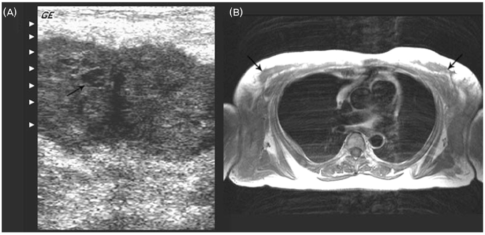
Figure 2 Ultrasonography and MRI of bilateral axillae. (A) Axial ultrasonography shows well-defined hypoechoic subcutaneous mass with internal echogenic content. (B) Axial T1-weighted image shows bilateral ill-defined subcutaneous masses in both axillae.
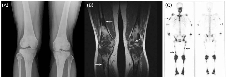
Figure 3 Radiological studies of bone. (A) A-P plain radiography of both knees show multifocal patchy areas of increased density, coarsened trabecullae, medullary sclerosis, and cortical thickening in symmetric distribution with involvement of diaphyses, metaphyses, and epiphyses. (B) Coronal T1-weighted MRI image shows hypointense lesions of geographic pattern at the diaphyses, metaphyses, and epiphyses. (C) Bone scan shows increased activity in the diaphyses, proximal and distal metaphyses, and ephiphyses of femur and tibia bilaterally and in the area of the right humerus.

Figure 4 Brain MRI. (A) T1-weighted axial MRI shows symmetrical hyperintense signals in the tegmentum pontis, right basis pontis, and right temporal lobe. (B) Gadolinium enhanced T1-weighted coronal MRI shows enhanced lesions in the right parietal lobe and middle cerebellar peduncle. (C) Gadolinium enhanced T1-weighted sagittal MRI shows moderate cerebellar atrophy and enhanced lesion in vermis part of the anterior lobe. (D) Gadolinium enhanced T1-weighted axial MRI shows an enhanced lesion in the right temporal lobe.
Reference
-
1. Veyssier-Belot C, Cacoub P, Caparros-Lefebvre D, Wechsler J, Brun B, Remy M, et al. Erdheim-Chester disease: Clinical and radiologic characteristics of 59 cases. Medicine. 1996. 75:157–169.
Article2. Chester W. Uber lipoidgranulomatose. Virchows Arch patholol Anat. 1930. 279:561–602.3. Clerico A, Ragni G, Cappelli C, Schiavetti A, Gonfiantini M, Uccini S. Erdheim-Chester disease in a child. Med Pediatr Oncol. 2003. 41:575–577.
Article4. Globerman H, Burstein S, Girardina PJ, Winchester P, Frankel S. A xanthogranulomatous histiocytosis in a child presenting with short stature. Am J Pediatr Hematol Oncol. 1991. 13:42–46.
Article5. Tan AP, Tan LK, Choo IH. Erdheim-Chester disease involving breast and muscle: imaging findings. AJR Am J Roentgenol. 1995. 164:1115–1117.
Article6. Lachenal F, Cotton F, Desmurs-Clavel H, Haroche J, Taillia H, Magy N, et al. Neurological manifestations and neuroradiological presentation of Erdheim-Chester disease: report of 6 cases and systematic review of the literature. J Neurol. 2006. 253:1267–1277.
Article7. Wright RA, Hermann RC, Parisi JE. Neurological manifestations of Erdheim-Chester disease. J Neurol Neurosurg Psychiatry. 1999. 66:72–75.
Article8. Weidauer S, von Stuckrad-Barre S, Dettmann E, Zanella FE, Lanfermann H. Cerebral Erdheim-Chester disease: case report and review of the literature. Neuroradiology. 2003. 45:241–245.
Article9. Grothe C, Urbach H, Bos M, Yo K, Schroder R. Cerebellar syndrome, exophthalmus and secondary hypogonadism in Erdheim-Chester disease. Nervenarzt. 2001. 72:449–452.10. Pautas E, Cherin P, Pelletier S, Vidailhet M, Herson S. Cerebral Erdheim-Chester disease: report of two cases with progressive cerebellar syndrome with dentate abnormalities on magnetic resonance imaging. J Neurol Neurosurg Psychiatry. 1998. 65:597–599.
Article11. Fukazawa T, Tsukishima E, Sasaki H, Hamada K, Hamada T, Tashiro K. Erdheim-Chester disease and slowly progressive cerebellar dysfunction. J Neurol Neurosurg Psychiatry. 1995. 58:238–240.
Article12. Kujat C, Junk B, Hermes M, Martin J, Dewes W. Cerebral manifestations of Erdheim-Chester disease. Radiologe. 1991. 31:307–309.13. Adle-Biassette H, Chetritt J, Bergemer-Fouquet AM, Wechsler J, Mussini JM, Gray F. Pathology of the central nervous system in Chester-Erdheim disease: report of three cases. J Neuropathol Exp Neurol. 1997. 56:1207–1216.14. Bohlega S, Alwatban J, Tulbah A, Bakheet SM, Powe J. Cerebral manifestation of Erdheim-Chester disease: clinical and radiologic findings. Neurology. 1997. 49:1702–1705.
Article15. Vital C, Bioulac-Sage P, Tison F, Rivel J, Begueret H, Gomez C, et al. Brain stem infiltration by mixed Langerhans cell histiocytosis and Erdheim-Chester disease: more than just an isolated case? Clin Exp Pathol. 1999. 47:71–76.
- Full Text Links
-
- Actions
-
Cited
- CITED
-
- Close
- Share
-
- Similar articles
-
- Erdheim-Chester Disease with Perirenal Masses Containing Macroscopic Fat Tissue
- Commentary on "A Case of Erdheim-Chester Disease with Asymptomatic Renal Involvement"
- Reply to Commentary on "A Case of Erdheim-Chester Disease with Asymptomatic Renal Involvement"
- A Case of Erdheim-Chester Disease with Bilateral Hydronephrosis
- Erdheim–Chester Disease Involving the Biliary System and Mimicking Immunoglobulin G4-Related Disease: A Case Report
