Erdheim-Chester Disease with Perirenal Masses Containing Macroscopic Fat Tissue
- Affiliations
-
- 1Department of Radiology, St. Vincent's Hospital, College of Medicine, The Catholic University of Korea, Suwon, Korea. bellenina@daum.net
- KMID: 2002813
- DOI: http://doi.org/10.3348/jksr.2015.72.2.143
Abstract
- Erdheim-Chester disease is a rare non-Langerhans-cell histiocytosis involving multiple organs. On histological evaluation, lipid-laden histiocyte aggregates in Erdheim-Chester disease is detected, but fat tissue in affected organs is not noted grossly on computed tomography. A 40-year-old man presented with bilateral perirenal masses containing fat tissue. He was diagnosed as perirenal involvement of Erdheim-Chester disease. This report describes a case of Erdheim-Chester disease with perirenal involvement that demonstrates unusual features.
MeSH Terms
Figure
-
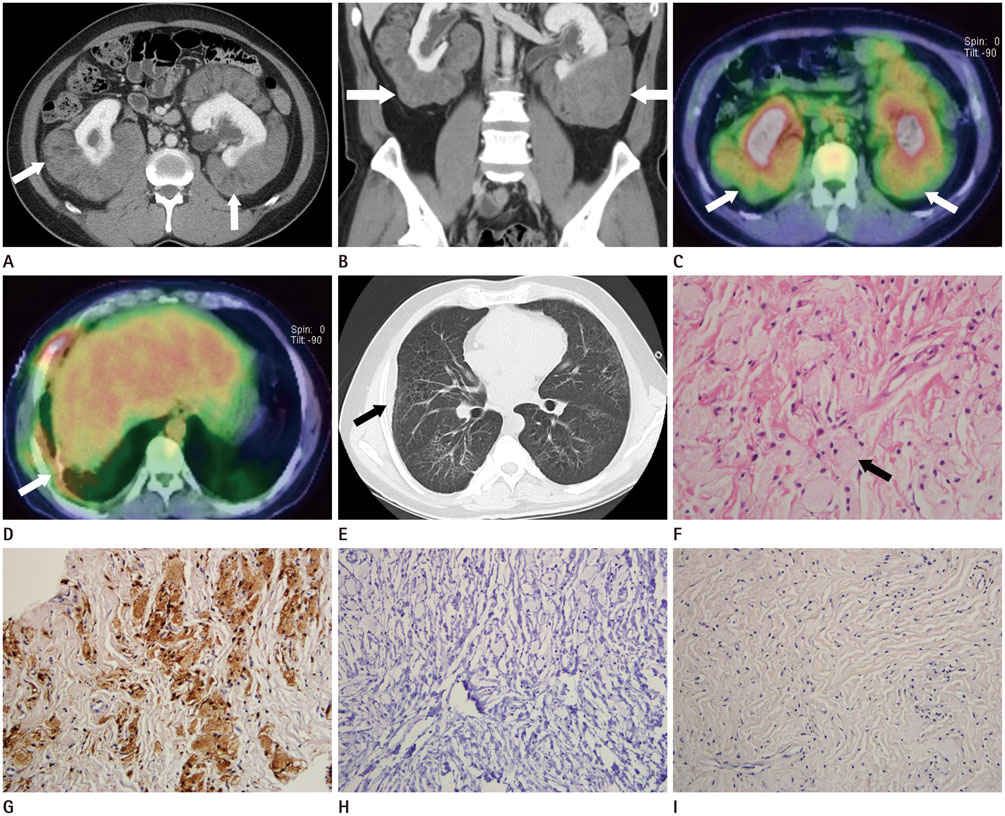
Fig. 1 A 40-year-old man with Erdheim-Chester disease. Bilateral perirenal masses with faint fat tissue (white arrows) are revealed on axial images (A) and coronal image of abdominal CT (B). Fused positron emission tomography (PET)/CT shows diffuse and moderate fluorodeoxyglucose (FDG) activity (maximum standardized uptake value range, 2.2-3.8) in bilateral perirenal masses (white arrows) (C). Some parts of lung and pleura (white arrow) also show mild patchy FDG activity on fused PET/CT (D). Multifocal ground glass opacities with interlobular and intralobular septal thickening and thickening of the right pleura (black arrow) are noted on chest CT (E). H&E stain (× 400) indicates lipid-laden histiocyte aggregation (black arrow for one among many lipid-laden histiocyte) (F). Immunohistochemical stains show that CD68 (× 200) (G) is positive and CD1a (× 200) (H) and S-100 (× 200) (I) are negative.
Reference
-
1. Alberti N, Frulio N, Bertolotti A, Petitpierre F, Veron A, Perez JT, et al. Erdheim-Chester disease: a rare diagnosis with evocative imaging. Diagn Interv Imaging. 2013; 94:457–459.2. Heller MT, Haarer KA, Thomas E, Thaete FL. Neoplastic and proliferative disorders of the perinephric space. Clin Radiol. 2012; 67:e31–e41.3. Dickson BC, Pethe V, Chung CT, Howarth DJ, Bilbao JM, Fornasier VL, et al. Systemic Erdheim-Chester disease. Virchows Arch. 2008; 452:221–227.4. De Filippo M, Ingegnoli A, Carloni A, Verardo E, Sverzellati N, Onniboni M, et al. Erdheim-Chester disease: clinical and radiological findings. Radiol Med. 2009; 114:1319–1329.5. Lee HJ, Lee KY, Shin DY, Lee YG, Choi SY, Moon KC, et al. A case of erdheim-chester disease with asymptomatic renal involvement. Cancer Res Treat. 2012; 44:146–150.6. Surabhi VR, Menias C, Prasad SR, Patel AH, Nagar A, Dalrymple NC. Neoplastic and non-neoplastic proliferative disorders of the perirenal space: cross-sectional imaging findings. Radiographics. 2008; 28:1005–1017.7. Provenzano E, Barter SJ, Wright PA, Forouhi P, Allibone R, Ellis IO. Erdheim-chester disease presenting as bilateral clinically malignant breast masses. Am J Surg Pathol. 2010; 34:584–588.8. Adam Z, Balsíková K, Krejcí M, Pour L, Stěpánková S, Svacina P, et al. [Central diabetes insipidus in adult patients--the first sign of Langerhans cell histiocytosis and Erdheim-Chester disease. Three case studies and literature review]. Vnitr Lek. 2010; 56:138–148.9. Adem C, Hélie O, Lévêque C, Taillia H, Cordoliani YS. Case 78: Erdheim-Chester disease with central nervous system involvement. Radiology. 2005; 234:111–115.
- Full Text Links
-
- Actions
-
Cited
- CITED
-
- Close
- Share
-
- Similar articles
-
- Commentary on "A Case of Erdheim-Chester Disease with Asymptomatic Renal Involvement"
- A case of Erdheim-Chester disease that presented with chronic renal failure
- A Case of Cerebral Erdheim-Chester Disease With Progressive Cerebellar Syndrome
- Reply to Commentary on "A Case of Erdheim-Chester Disease with Asymptomatic Renal Involvement"
- Erdheim-Chester Disease
