Erdheim-Chester Disease
- Affiliations
-
- 1Department of Dermatology, Asan Medical Center, University of Ulsan College of Medicine, Seoul, Korea. miumiu@amc.seoul.kr
- KMID: 2266183
- DOI: http://doi.org/10.5021/ad.2010.22.4.439
Abstract
- Erdheim-Chester disease (ECD) is a rare, non-Langerhans cell histiocytosis of unknown etiology, characterized by multi-organ involvement. ECD is usually diagnosed on the basis of characteristic radiologic and histopathological findings. Lesions may be skeletal and/or extraskeletal in location, and may include the skin, lung, heart, and central nervous system. We describe here a 68-year-old man with multiple yellowish plaques and a pinkish nodule on his face and scalp. He had been previously diagnosed with diabetes insipidus, and recently complained of coughing and dyspnea. Imaging studies showed multiple osteosclerotic lesions of the bones, a moderate amount of pericardial effusion, and multifocal infiltrative lesions in the perirenal space. Histopathological examination of the skin lesions revealed dermal infiltration of foamy histiocytes with multinuclear giant cells. Moreover, laparoscopic biopsy of the perirenal tissue revealed fibrosis with infiltrating foamy histiocytes being CD68-positive and S100-negative. Based on these findings, he was diagnosed with ECD with extraskeletal manifestations, and treated with interferon-alpha.
MeSH Terms
Figure
-

Fig. 1 (A, B) Multiple and slightly-raised yellowish plaques on the left temple, forehead and both lower eyelids. (C) A pinkish dome-shaped nodule (approximately 1 cm) on the parietal scalp.

Fig. 2 (A) Left temporal lesion showed dermal infiltration of foamy histiocytes with giant cells (H&E, ×100). (B) Scalp lesion also showed infiltrating histiocytes and multiple Touton-type giant cells with dermal fibrosis (H&E, ×100).
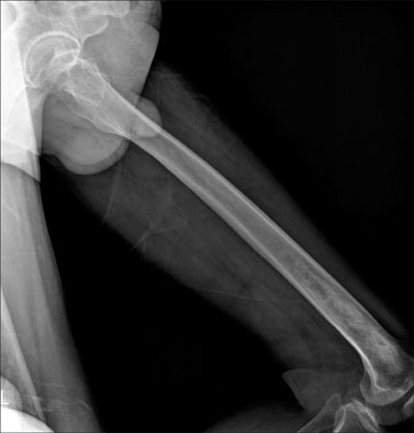
Fig. 3 X-ray of left femur revealed diffuse osteosclerosis with focal osteolysis in the diaphysis and metaphysis of the left distal femur.
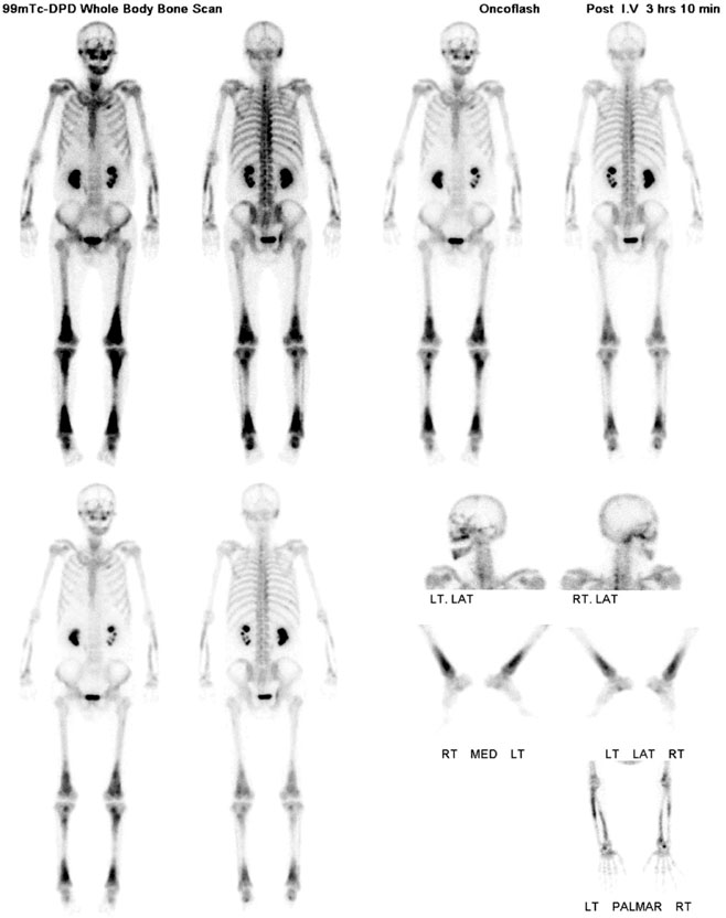
Fig. 4 Bone scan showed multiple increased uptakes in both maxillae, mandible, right temporal bone, both radii, both ulnae, both distal femurs, both proximal and distal tibiae, both calcanei, left second rib, right ninth rib, both tenth ribs, left posterior iliac crest and right acetabulum.

Fig. 5 (A) The biopsy obtained from perirenal tissue showed an infiltration of numerous histiocytes (H&E, ×100). The histiocytes were CD68-positive (B: ×100) and S100-negative (C: ×100).
Reference
-
1. Veyssier-Belot C, Cacoub P, Caparros-Lefebvre D, Wechsler J, Brun B, Remy M, et al. Erdheim-Chester disease. Clinical and radiologic characteristics of 59 cases. Medicine (Baltimore). 1996. 75:157–169.
Article2. Sheu SY, Wenzel RR, Kersting C, Merten R, Otterbach F, Schmid KW. Erdheim-Chester disease: case report with multisystemic manifestations including testes, thyroid, and lymph nodes, and a review of literature. J Clin Pathol. 2004. 57:1225–1228.
Article3. Myra C, Sloper L, Tighe PJ, McIntosh RS, Stevens SE, Gregson RH, et al. Treatment of Erdheim-Chester disease with cladribine: a rational approach. Br J Ophthalmol. 2004. 88:844–847.
Article4. Dickson BC, Pethe V, Chung CT, Howarth DJ, Bilbao JM, Fornasier VL, et al. Systemic Erdheim-Chester disease. Virchows Arch. 2008. 452:221–227.
Article5. Greenberger JS, Crocker AC, Vawter G, Jaffe N, Cassady JR. Results of treatment of 127 patients with systemic histiocytosis. Medicine (Baltimore). 1981. 60:311–338.6. Suzuki HI, Hosoya N, Miyagawa K, Ota S, Nakashima H, Makita N, et al. Erdheim-Chester disease: multisystem involvement and management with interferon-alpha. Leuk Res. 2010. 34:e21–e24.7. Breuil V, Brocq O, Pellegrino C, Grimaud A, Euller-Ziegler L. Erdheim-Chester disease: typical radiological bone features for a rare xanthogranulomatosis. Ann Rheum Dis. 2002. 61:199–200.
Article8. Perlat A, Decaux O, Sébillot M, Grosbois B, Desfourneaux V, Meadeb J. Erdheim-Chester disease with predominant mesenteric localization: lack of efficacy of interferon alpha. Joint Bone Spine. 2009. 76:315–317.
Article9. Haroche J, Amoura Z, Trad SG, Wechsler B, Cluzel P, Grenier PA, et al. Variability in the efficacy of interferon-alpha in Erdheim-Chester disease by patient and site of involvement: results in eight patients. Arthritis Rheum. 2006. 54:3330–3336.
Article
- Full Text Links
-
- Actions
-
Cited
- CITED
-
- Close
- Share
-
- Similar articles
-
- Erdheim-Chester Disease with Perirenal Masses Containing Macroscopic Fat Tissue
- Commentary on "A Case of Erdheim-Chester Disease with Asymptomatic Renal Involvement"
- Reply to Commentary on "A Case of Erdheim-Chester Disease with Asymptomatic Renal Involvement"
- A Case of Erdheim-Chester Disease with Bilateral Hydronephrosis
- Intracranial Erdheim-Chester Disease
