J Clin Neurol.
2014 Oct;10(4):363-366. 10.3988/jcn.2014.10.4.363.
Hydrocephalus and Neurocysticercosis: Cases Illustrative of Three Distinct Mechanisms
- Affiliations
-
- 1Department of Neurosurgery, AP-HP Hopital Beaujon, Clichy, France. aymmed@hotmail.fr
- KMID: 2287518
- DOI: http://doi.org/10.3988/jcn.2014.10.4.363
Abstract
- BACKGROUND
Cysticercosis is the most frequent parasitic infection of the nervous system. Most lesions are intracranial, and spinal involvement is rare. We describe here in two cases of neurocysticercosis (NCC) in the brain and one in the spinal cord that illustrate three distinct mechanisms leading to symptomatic acute hydrocephalus.
CASE REPORT
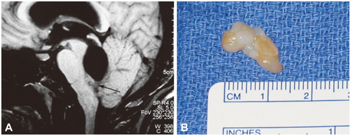
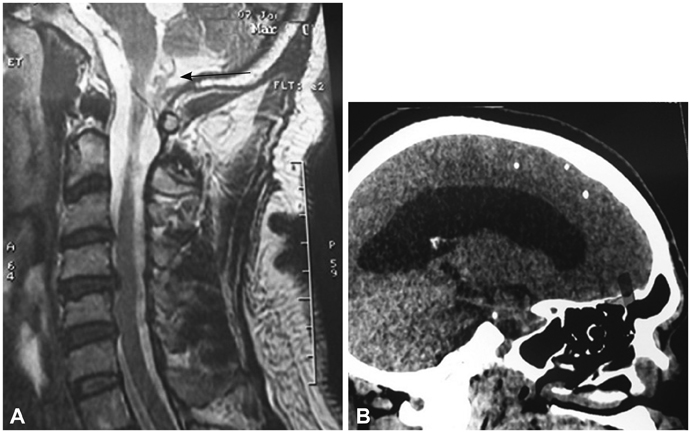
Hydrocephalus was related to intracranial NCC in two of them. In the first case the hydrocephalus was due to an extensive arachnoiditis to the craniocervical junction, while in the second it was caused by obstruction of Magendie's foramen in the fourth ventricle by the scolex of Taenia solium. For the third patient, hydrocephalus revealed cysticercosis of the cauda equina due to the scolex.
CONCLUSIONS
NCC should be considered as a possible diagnosis for patients suffering from hydrocephalus when they originate from or have traveled in endemic areas, MRI of the spine is mandatory to search for intraspinal lesions.
Keyword
MeSH Terms
Figure
Reference
-
1. Román G, Sotelo J, Del Brutto O, Flisser A, Dumas M, Wadia N, et al. A proposal to declare neurocysticercosis an international reportable disease. Bull World Health Organ. 2000; 78:399–406.2. Lobato RD, Lamas E, Portillo JM, Roger R, Esparza J, Rivas JJ, et al. Hydrocephalus in cerebral cysticercosis. Pathogenic and therapeutic considerations. J Neurosurg. 1981; 55:786–793.3. Raccurt CP, Agnamey P, Boncy J, Henrys JH, Totet A. Seroprevalence of human Taenia solium cysticercosis in Haiti. J Helminthol. 2009; 83:113–116.
Article4. Bouree P, Dumazedier D, Bisaro F, Resende P, Comoy J, Aghakhani N. Spinal cord cysticercosis: a case report. J Egypt Soc Parasitol. 2006; 36:727–736.5. Monteiro L, Almeida-Pinto J, Stocker A, Sampaio-Silva M. Active neurocysticercosis, parenchymal and extraparenchymal: a study of 38 patients. J Neurol. 1993; 241:15–21.
Article6. Matushita H, Pinto FC, Cardeal DD, Teixeira MJ. Hydrocephalus in neurocysticercosis. Childs Nerv Syst. 2011; 27:1709–1721.
Article7. Salazar A, Sotelo J, Martinez H, Escobedo F. Differential diagnosis between ventriculitis and fourth ventricle cyst in neurocysticercosis. J Neurosurg. 1983; 59:660–663.
Article8. Apuzzo ML, Dobkin WR, Zee CS, Chan JC, Giannotta SL, Weiss MH. Surgical considerations in treatment of intraventricular cysticercosis. An analysis of 45 cases. J Neurosurg. 1984; 60:400–407.9. Sotelo J, del Brutto OH, Penagos P, Escobedo F, Torres B, Rodriguez-Carbajal J, et al. Comparison of therapeutic regimen of anticysticercal drugs for parenchymal brain cysticercosis. J Neurol. 1990; 237:69–72.
Article10. Ahmad FU, Sharma BS. Treatment of intramedullary spinal cysticercosis: report of 2 cases and review of literature. Surg Neurol. 2007; 67:74–77. discussion 77.
Article11. Zee CS, Segall HD, Destian S, Ahmadi J, Apuzzo ML. MRI of intraventricular cysticercosis: surgical implications. J Comput Assist Tomogr. 1993; 17:932–939.12. Mirone G, Cinalli G, Spennato P, Ruggiero C, Aliberti F. Hydrocephalus and spinal cord tumors: a review. Childs Nerv Syst. 2011; 27:1741–1749.
Article13. Agrawal R, Chauhan SP, Misra V, Singh PA, Gopal NN. Focal spinal intramedullary cysticercosis. Acta Biomed. 2008; 79:39–41.14. Gardner WJ, Spitler DK, Whitten C. Increased intracranial pressure caused by increased protein content in the cerebrospinal fluid; an explanation of papilledema in certain cases of small intracranial and intraspinal tumors, and in the Guillain-Barre syndrome. N Engl J Med. 1954; 250:932–936.
Article15. Morandi X, Amlashi SF, Riffaud L. A dynamic theory for hydrocephalus revealing benign intraspinal tumours: tumoural obstruction of the spinal subarachnoid space reduces total CSF compartment compliance. Med Hypotheses. 2006; 67:79–81.
Article16. Raksin PB, Alperin N, Sivaramakrishnan A, Surapaneni S, Lichtor T. Noninvasive intracranial compliance and pressure based on dynamic magnetic resonance imaging of blood flow and cerebrospinal fluid flow: review of principles, implementation, and other noninvasive approaches. Neurosurg Focus. 2003; 14:e4.
Article17. Garcia HH, Rodriguez S, Gilman RH, Gonzalez AE, Tsang VC. Cysticercosis Working Group in Peru. Neurocysticercosis: is serology useful in the absence of brain imaging? Trop Med Int Health. 2012; 17:1014–1018.
Article
- Full Text Links
- Actions
-
Cited
- CITED
-
- Close
- Share
- Similar articles
-
- Calcified Neurocysticercosis that Invaded the Subarachnoid Space Presenting as Focal Status Epilepticus
- Transaqueductal Migration of the Neurocysticercus Cyst: Two Case Report
- Neurocysticercosis-New Aspects of Diagnosis and Management after the Introduction of ELISA and Praziquantel
- Complications after the Ventriculo-peritoneal Shunt according to the Time Course
- Acquired Chiari-malformation after Ventriculoperitoneal Shunt for Hydrocepalus Associated with Neurocysticercosis


