Malignant Fibrous Histiocytoma in the Breast: A Case Report
- Affiliations
-
- 1Department of Surgery, The Catholic University of Korea, College of Medicine, Seoul, Korea. gsshchoi@catholic.ac.kr
- 2Department of Clinical Pathology, The Catholic University of Korea, College of Medicine, Seoul, Korea.
- 3Department of Radiology, The Catholic University of Korea, College of Medicine, Seoul, Korea.
- KMID: 2286588
- DOI: http://doi.org/10.4048/jbc.2008.11.4.213
Abstract
- Malignant fibrous histiocytoma is the most common form of soft tissue sarcoma during middle and late adulthood in the deep connective tissues of the extremities, abdominal cavity, and retroperitoneum. Primary breast sarcoma is a rare disease entity, comprising less than 1% of all breast malignancies. Malignant fibrous histiocytoma of the breast is very rare. We presented one case of a malignant fibrous histiocytoma of the right breast in a 49-year-old woman and report the case with a review of the literature.
Keyword
MeSH Terms
Figure
-
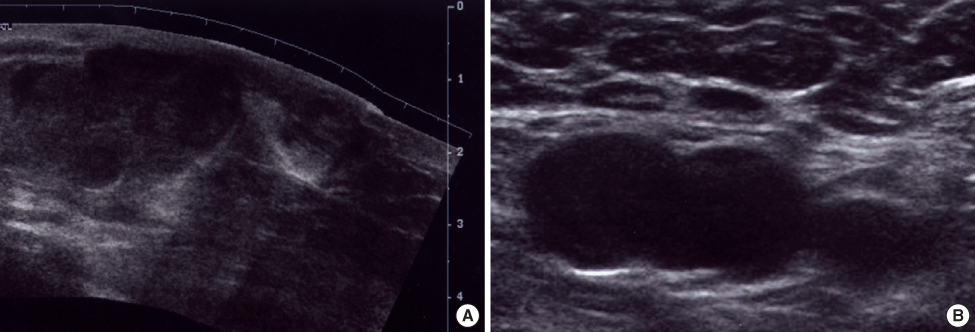
Fig 1 The ultrasonographic findings of malignant fibrous histiocytoma. Several large hypoechoic & isoechoic masses are noted in lower half of right breast and part of them are located in subcutaneous layer (A) and lymph nodes in right axilla are enlarged (B). These findings are included in BI-RADS category 5.
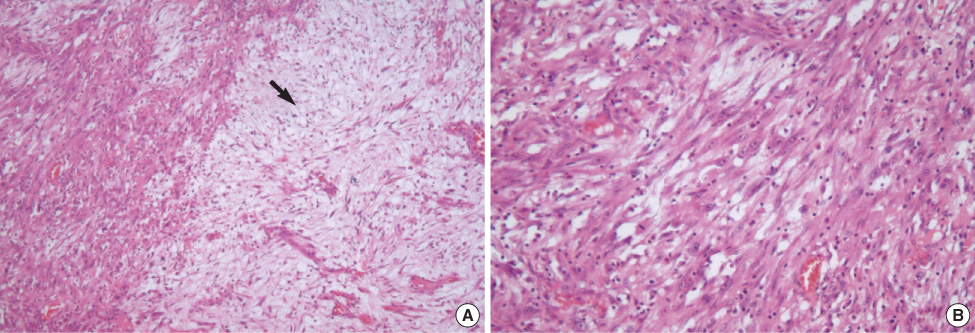
Fig 2 Histopathologic findings of malignant fibrous histiocytoma (A & B; ×40 & ×100, HE stain) Highly pleomorphic spindle shaped cells are observed in cellular tumor tissue. The tumor shows broad myxoid tissue (arrow) abutting cellular area (A) and fascicular pattern of pleomorphic spindle shaped cells (B).
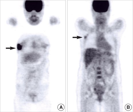
Fig 3 PET-CT findings. Intense F-18 FDG accumulation (arrow) in subareolar and lower lateral portion of right breast (A) and several focal FDG uptake (arrow) in right axilla (B) are observed.
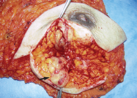
Fig 4 The mastectomy specimen. A relatively well circumscribed ovoid mass (arrow) is located beneath skin, which is 3.0×2.5 cm in size and has yellowish to pale gray myxoid and soft cut surface with focal hemorrhage.
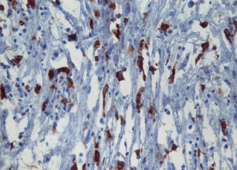
Fig 5 Immunohistochemical staining of CD68 (×400). The tumor is positive for CD68.
Reference
-
1. Yao MS, Chan WP, Chen AY, Chu JS, Hsieh MC. Malignant fibrous histiocytoma of the female breast: a case report. Clin Imaging. 2005. 29:134–137.2. Blanchard DK, Reynolds CA, Grant CS, Donohue JH. Primary nonphylloides breast sarcomas. Am J Surg. 2003. 186:359–361.
Article3. Tamir G, Nobel M, Hauben DJ, Sandbank J. Malignant fibrous histiocytoma of the breast. Eur J Surg Oncol. 1995. 21:210–211.
Article4. Doussal VL, Coindre JM, Leroux A, Hacene K, Terrier P, Bui NB, et al. Prognostic factors for patients with localized primary malignant fibrous hiostiocytoma; a multicenter study of 216 patients with multivariate analysis. Cancer. 1996. 77:1823–1830.
Article5. Matsumoto S, Ahmed AR, Kawaguchi N, Manabe J, Marsushita Y. Results of surgery for malignant fibrous histiocytomas of soft tissue. Int J Clin Oncol. 2003. 8:104–109.
Article6. Oh SJ, Kim KM, Hong TH, Park WC, Kim JS, Jung SS. Giant cell malignant fibrous hiotiocytoma of the breast: a case report. J Korean Med Sci. 2004. 19:477–480.
Article7. Berg JW, DeCosse JJ, Fracchia AA, Farrow JH. Stromal sarcomas of the breast. A unified approach to connective sarcomas other than cystosarcoma phyllodes. Cancer. 1962. 15:418–424.
Article8. Callery CD, Rosen PP, Kinne DW. Sarcoma of the breast. A study of 32 patients with reappraisal of classification and therapy. Ann Surg. 1985. 201:527–532.9. O'Brien JE, Stout AP. Malignant fibrous xanthomas. Cancer. 1964. 17:1445–1455.10. Langhan MR, Mills AS, De May RM, O'Dowd GJ, Grathwohl MA, Horsley JS. Malignant fibrous histiocytoma of the breast. A case report and review of the literature. Cancer. 1984. 54:558–563.
Article11. Jones MW, Norris HJ, Wargotz ES, Weiss SW. Fibrosarcoma-malignant fibrous histiocytoma of the breast. A clinicopathological study of 32 cases. Am J surg Pathol. 1992. 16:667–674.12. Weiss SW. Malignant fibrous histiocytoma. A reaffirmation. Am J Surg Pathol. 1982. 6:773–784.13. Wiriosuparto S, Krassilnik N, Gologan A, Cohen JM, Wenig B. Malignant fibrous histiocytoma, giant cell type, of the breast mimicking metaplastic carcinoma. Acta Cytol. 2003. 47:673–679.
Article14. McGowan TS, Cummings BJ, O'Sullivan B, Catton CN, Miller N, Panjarella T. An analysis of 78 breast sarcoma patients without distant metastases at presentation. Int J Radiot Oncol Biol Phys. 2000. 46:383–390.
Article15. American Joint Committee on Cancer. AJCC Cancer Staging Manual. 2002. 6th ed. NewYork: Springer-Verlag.
- Full Text Links
-
- Actions
-
Cited
- CITED
-
- Close
- Share
-
- Similar articles
-
- A Case of the retroperitoneal Malignant Fibrous Histiocytoma
- Malignant Fibrous Histiocytoma: A Case Report
- Malignant fibrous histiocytoma
- Post-Radiation Malignant Fibrous Histiocytoma Following Treatment of Breast Cancer: A Case Report with Imaging Findings
- Retroperitoneal Malignant Fibrous Histiocytoma: Report of a Case
