Post-Radiation Malignant Fibrous Histiocytoma Following Treatment of Breast Cancer: A Case Report with Imaging Findings
- Affiliations
-
- 1Department of Surgery, Dankook University College of Medicine, Seoul, Korea.
- 2Department of Pathology, Dankook University College of Medicine, Seoul, Korea.
- 3Department of Radiology, Dankook University College of Medicine, Seoul, Korea. sirenos@hanmail.net
- KMID: 2041952
- DOI: http://doi.org/10.3348/jksr.2013.69.4.307
Abstract
- Post-radiation malignant fibrous histiocytoma (MFH) of the breast is extremely rare. We report a case of post-radiation MFH that presented a rapidly growing mass in a 52-year-old woman who underwent breast-conserving therapy and adjuvant whole breast irradiation 6 years ago. To the best of our knowledge, only one case of primary MFH of the female breast have been reported with sonographic findings. We analyzed the sonographic and MRI findings with correlative histopathologic features, and then confirmed with surgical excision.
Figure
-
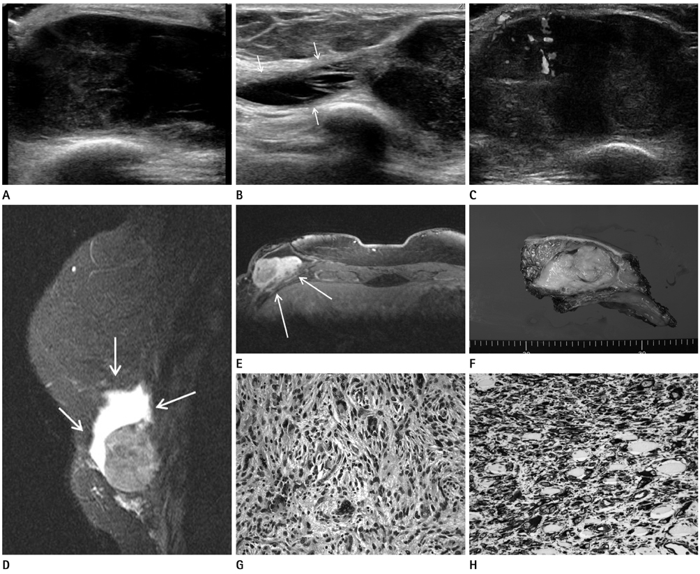
Fig. 1 A 52-year-old woman with a rapidly growing mass in her right lower breast near the inframammary fold. A. Ultrasonography reveal a 6 × 2.8 cm oval, circumscribed mass with a complex echogenicity at the 6 o'clock position. B. Elongated septated cystic area can be seen at the upper portion of mass (arrows). C. On color Doppler sonography, there is some vascular flow within the mass. D. A sagittal, fat-saturated, T2-weighted MR image shows an approximately 6 cm mass with high signal intensity, and peripheral portion of the mass showed avidly high signal intensity, which suggested cystic area (arrows). E. A post-contrast fat-suppressed T1 weighted MR axial image demonstrate a inhomogeneous enhancing solid tumor with surrounding non-enhancing cystic area invading the pectoralis major muscle at the right lower inframammary fold area (arrows). F. A yellow solid tumor was totally excised along with the pectoralis muscle and ribs. G. Microscopic features consisting of plump spindle cells and pleomorphic cells with mitotic activity (hematoxyline and eosin stain, original magnification × 200). H. Immunohistochemical staining demonstrating diffuse cytoplasmic positivity of tumor cells (Immunohistochemistry, original magnification × 200).
Reference
-
1. Kirova YM, Vilcoq JR, Asselain B, Sastre-Garau X, Fourquet A. Radiation-induced sarcomas after radiotherapy for breast carcinoma: a large-scale single-institution review. Cancer. 2005; 104:856–863.2. Fisher B, Anderson S, Bryant J, Margolese RG, Deutsch M, Fisher ER, et al. Twenty-year follow-up of a randomized trial comparing total mastectomy, lumpectomy, and lumpectomy plus irradiation for the treatment of invasive breast cancer. N Engl J Med. 2002; 347:1233–1241.3. Mery CM, George S, Bertagnolli MM, Raut CP. Secondary sarcomas after radiotherapy for breast cancer: sustained risk and poor survival. Cancer. 2009; 115:4055–4063.4. Biswas S, Badiuddin F. Radiation induced malignant histiocytoma of the contralateral breast following treatment of breast cancer: a case report and review of the literature. Cases J. 2008; 1:313.5. Yap J, Chuba PJ, Thomas R, Aref A, Lucas D, Severson RK, et al. Sarcoma as a second malignancy after treatment for breast cancer. Int J Radiat Oncol Biol Phys. 2002; 52:1231–1237.6. Pendlebury SC, Bilous M, Langlands AO. Sarcomas following radiation therapy for breast cancer: a report of three cases and a review of the literature. Int J Radiat Oncol Biol Phys. 1995; 31:405–410.7. Sheppard DG, Libshitz HI. Post-radiation sarcomas: a review of the clinical and imaging features in 63 cases. Clin Radiol. 2001; 56:22–29.8. Sheth GR, Cranmer LD, Smith BD, Grasso-Lebeau L, Lang JE. Radiation-induced sarcoma of the breast: a systematic review. Oncologist. 2012; 17:405–418.9. Yao MS, Chan WP, Chen CY, Chu JS, Hsieh MC. Malignant fibrous histiocytoma of the female breast: a case report. Clin Imaging. 2005; 29:134–137.10. Noh JM, Huh SJ, Choi DH, Park W, Nam SJ. Two cases of post-radiation sarcoma after breast cancer treatment. J Breast Cancer. 2012; 15:364–370.
- Full Text Links
-
- Actions
-
Cited
- CITED
-
- Close
- Share
-
- Similar articles
-
- Imaging Findings of Metastatic Breast Malignant Fibrous Histiocytoma: A Case Report
- Malignant Fibrous Histiocytoma: A Case Report
- A Case Report of Primary Mediastinal Malignant Fibrous Histiocytoma
- A Case of the retroperitoneal Malignant Fibrous Histiocytoma
- Imaging Findings of Malignant Fibrous Histiocytoma of the Breast: A Case Report
