Korean J Physiol Pharmacol.
2016 May;20(3):269-277. 10.4196/kjpp.2016.20.3.269.
Ellagic acid plays a protective role against UV-B-induced oxidative stress by up-regulating antioxidant components in human dermal fibroblasts
- Affiliations
-
- 1Department of Biochemistry, College of Natural Sciences, Kangwon National University, Chuncheon 24341, Korea. cjlim@kangwon.ac.kr
- 2Department of Biological Sciences, College of Natural Sciences, Kangwon National University, Chuncheon 24341, Korea. kkim@kangwon.ac.kr
- 3Shebah Biotech Inc., Chuncheon 24398, Korea.
- KMID: 2285599
- DOI: http://doi.org/10.4196/kjpp.2016.20.3.269
Abstract
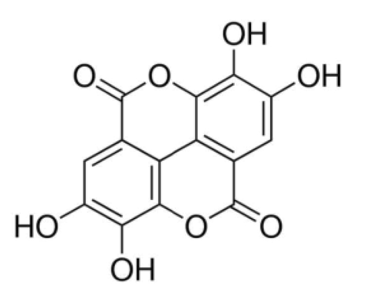
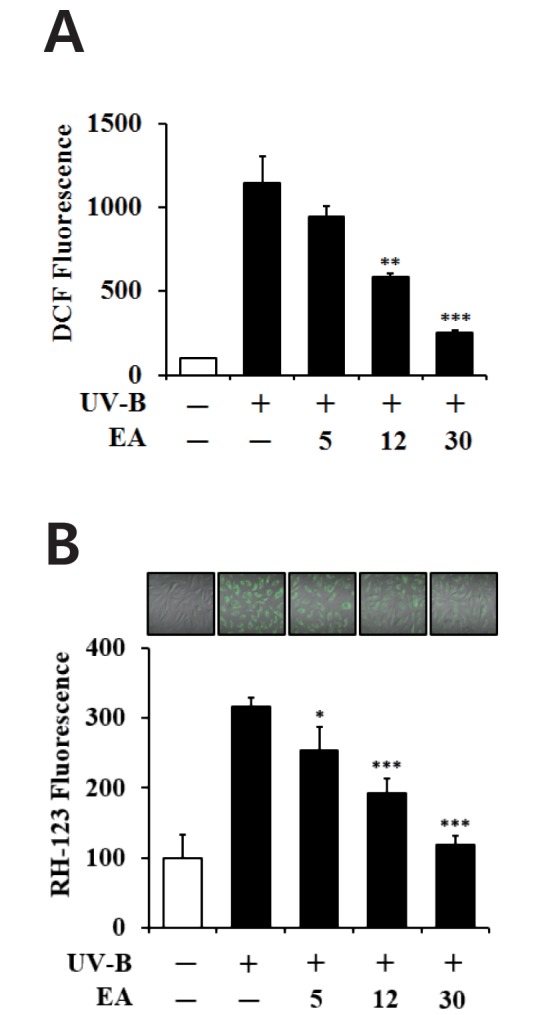
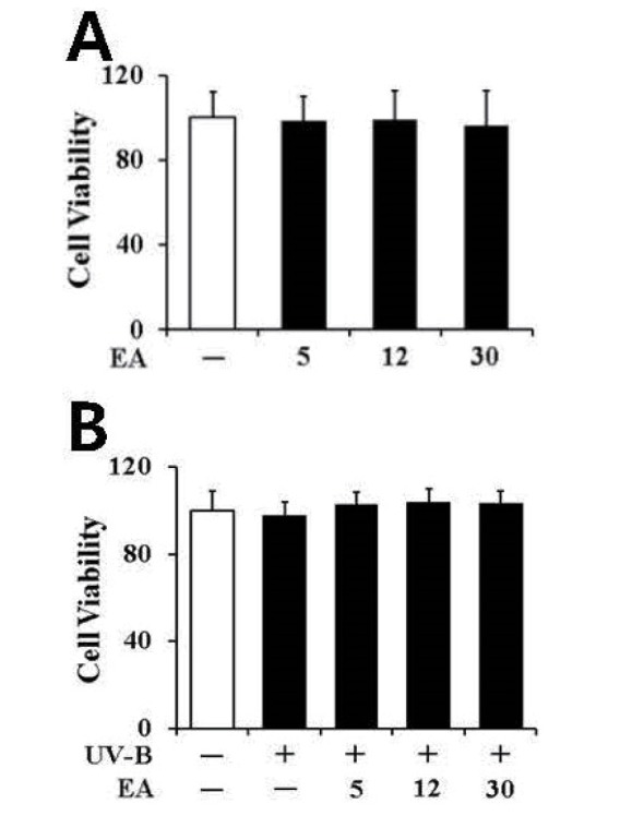
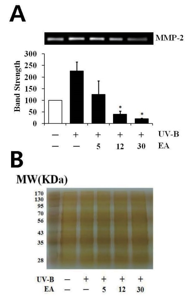
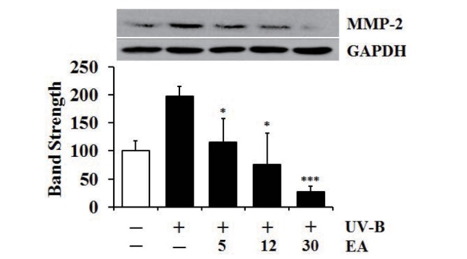
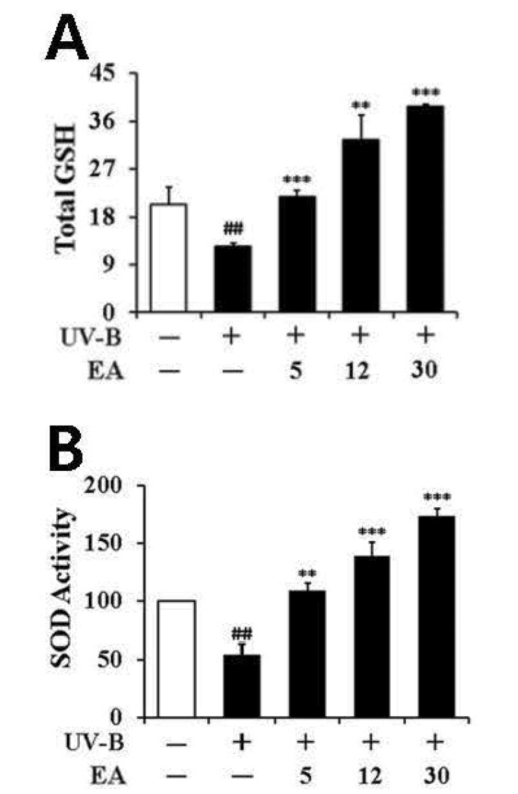
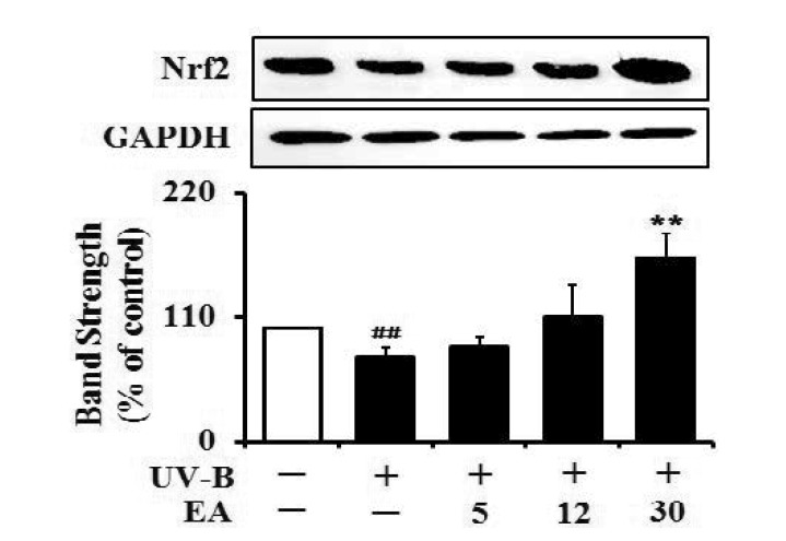
- Ellagic acid (EA), an antioxidant polyphenolic constituent of plant origin, has been reported to possess diverse pharmacological properties, including anti-inflammatory, anti-tumor and immunomodulatory activities. This work aimed to clarify the skin anti-photoaging properties of EA in human dermal fibroblasts. The skin anti-photoaging activity was evaluated by analyzing the reactive oxygen species (ROS), matrix metalloproteinase-2 (MMP-2), total glutathione (GSH) and superoxide dismutase (SOD) activity levels as well as cell viability in dermal fibroblasts under UV-B irradiation. When fibroblasts were exposed to EA prior to UV-B irradiation, EA suppressed UV-B-induced ROS and proMMP-2 elevation. However, EA restored total GSH and SOD activity levels diminished in fibroblasts under UV-B irradiation. EA had an up-regulating activity on the UV-B-reduced Nrf2 levels in fibroblasts. EA, at the concentrations used, was unable to interfere with cell viabilities in both non-irradiated and irradiated fibroblasts. In human dermal fibroblasts, EA plays a defensive role against UV-B-induced oxidative stress possibly through an Nrf2-dependent pathway, indicating that this compound has potential skin antiphotoaging properties.
MeSH Terms
Figure
Reference
-
1. Galano A, Francisco Marquez M, Pérez-González A. Ellagic acid: an unusually versatile protector against oxidative stress. Chem Res Toxicol. 2014; 27:904–918. PMID: 24697747.
Article2. Larrosa M, Tomás-Barberán FA, Espín JC. The dietary hydrolysable tannin punicalagin releases ellagic acid that induces apoptosis in human colon adenocarcinoma Caco-2 cells by using the mitochondrial pathway. J Nutr Biochem. 2006; 17:611–625. PMID: 16426830.
Article3. Goodwin EC, Atwood WJ, DiMaio D. High-throughput cell-based screen for chemicals that inhibit infection by simian virus 40 and human polyomaviruses. J Virol. 2009; 83:5630–5639. PMID: 19297472.
Article4. Rogerio AP, Fontanari C, Borducchi E, Keller AC, Russo M, Soares EG, Albuquerque DA, Faccioli LH. Anti-inflammatory effects of Lafoensia pacari and ellagic acid in a murine model of asthma. Eur J Pharmacol. 2008; 580:262–270. PMID: 18021768.
Article5. Seeram NP, Adams LS, Henning SM, Niu Y, Zhang Y, Nair MG, Heber D. In vitro antiproliferative, apoptotic and antioxidant activities of punicalagin, ellagic acid and a total pomegranate tannin extract are enhanced in combination with other polyphenols as found in pomegranate juice. J Nutr Biochem. 2005; 16:360–367. PMID: 15936648.
Article6. Pari L, Sivasankari R. Effect of ellagic acid on cyclosporine A-induced oxidative damage in the liver of rats. Fundam Clin Pharmacol. 2008; 22:395–401. PMID: 18705750.
Article7. Han DH, Lee MJ, Kim JH. Antioxidant and apoptosis-inducing activities of ellagic acid. Anticancer Res. 2006; 26:3601–3606. PMID: 17094489.8. Kilic I, Yeşiloğlu Y, Bayrak Y. Spectroscopic studies on the antioxidant activity of ellagic acid. Spectrochim Acta A Mol Biomol Spectrosc. 2014; 130:447–452. PMID: 24813273.
Article9. Birch-Machin MA, Swalwell H. How mitochondria record the effects of UV exposure and oxidative stress using human skin as a model tissue. Mutagenesis. 2010; 25:101–107. PMID: 19955330.
Article10. Barresi C, Stremnitzer C, Mlitz V, Kezic S, Kammeyer A, Ghannadan M, Posa-Markaryan K, Selden C, Tschachler E, Eckhart L. Increased sensitivity of histidinemic mice to UVB radiation suggests a crucial role of endogenous urocanic acid in photoprotection. J Invest Dermatol. 2011; 131:188–194. PMID: 20686493.
Article11. Ikehata H, Ono T. The mechanisms of UV mutagenesis. J Radiat Res. 2011; 52:115–125. PMID: 21436607.
Article12. Peres PS, Terra VA, Guarnier FA, Cecchini R, Cecchini AL. Photoaging and chronological aging profile: Understanding oxidation of the skin. J Photochem Photobiol B. 2011; 103:93–97. PMID: 21356598.
Article13. Curran S, Murray GI. Matrix metalloproteinases in tumour invasion and metastasis. J Pathol. 1999; 189:300–308. PMID: 10547590.
Article14. Pygmalion MJ, Ruiz L, Popovic E, Gizard J, Portes P, Marat X, Lucet-Levannier K, Muller B, Galey JB. Skin cell protection against UVA by Sideroxyl, a new antioxidant complementary to sunscreens. Free Radic Biol Med. 2010; 49:1629–1637. PMID: 20826208.
Article15. Lee YM, Kim YK, Kim KH, Park SJ, Kim SJ, Chung JH. A novel role for the TRPV1 channel in UV-induced matrix metalloproteinase (MMP)-1 expression in HaCaT cells. J Cell Physiol. 2009; 219:766–775. PMID: 19206161.
Article16. Chae S, Piao MJ, Kang KA, Zhang R, Kim KC, Youn UJ, Nam KW, Lee JH, Hyun JW. Inhibition of matrix metalloproteinase-1 induced by oxidative stress in human keratinocytes by mangiferin isolated from Anemarrhena asphodeloides. Biosci Biotechnol Biochem. 2011; 75:2321–2325. PMID: 22146720.17. Kim MS, Oh GH, Kim MJ, Hwang JK. Fucosterol inhibits matrix metalloproteinase expression and promotes type-1 procollagen production in UVB-induced HaCaT cells. Photochem Photobiol. 2013; 89:911–918. PMID: 23418792.
Article18. Hseu YC, Chou CW, Senthil Kumar KJ, Fu KT, Wang HM, Hsu LS, Kuo YH, Wu CR, Chen SC, Yang HL. Ellagic acid protects human keratinocyte (HaCaT) cells against UVA-induced oxidative stress and apoptosis through the upregulation of the HO-1 and Nrf-2 antioxidant genes. Food Chem Toxicol. 2012; 50:1245–1255. PMID: 22386815.
Article19. Lembo S, Balato A, Di Caprio R, Cirillo T, Giannini V, Gasparri F, Monfrecola G. The modulatory effect of ellagic acid and rosmarinic acid on ultraviolet-B-induced cytokine/chemokine gene expression in skin keratinocyte (HaCaT) cells. Biomed Res Int. 2014; DOI: 10.1155/2014/346793.
Article20. Bradford MM. A rapid and sensitive method for the quantitation of microgram quantities of protein utilizing the principle of proteindye binding. Anal Biochem. 1976; 72:248–254. PMID: 942051.
Article21. Royall JA, Ischiropoulos H. Evaluation of 2',7'-dichlorofluorescin and dihydrorhodamine 123 as fluorescent probes for intracellular H2O2 in cultured endothelial cells. Arch Biochem Biophys. 1993; 302:348–355. PMID: 8387741.
Article22. Freshney RI. Culture of animal cells: a manual of basic technique. 3rd ed. New York: Wiley-Liss Press;1994.23. Kleiner DE, Stetler-Stevenson WG. Quantitative zymography: detection of picogram quantities of gelatinases. Anal Biochem. 1994; 218:325–329. PMID: 8074288.
Article24. Nakagawa K, Saijo N, Tsuchida S, Sakai M, Tsunokawa Y, Yokota J, Muramatsu M, Sato K, Terada M, Tew KD. Glutathione-S-transferase pi as a determinant of drug resistance in transfectant cell lines. J Biol Chem. 1990; 265:4296–4301. PMID: 2407735.
Article25. Lee YY, Kim HG, Jung HI, Shin YH, Hong SM, Park EH, Sa JH, Lim CJ. Activities of antioxidant and redox enzymes in human normal hepatic and hepatoma cell lines. Mol Cells. 2002; 14:305–311. PMID: 12442906.26. Quan T, Qin Z, Xia W, Shao Y, Voorhees JJ, Fisher GJ. Matrix-degrading metalloproteinases in photoaging. J Investig Dermatol Symp Proc. 2009; 14:20–24.
Article27. Zhu M, Bowden GT. Molecular mechanism(s) for UV-B irradiation-induced glutathione depletion in cultured human keratinocytes. Photochem Photobiol. 2004; 80:191–196. PMID: 15244506.
Article28. Priyadarsini KI, Khopde SM, Kumar SS, Mohan H. Free radical studies of ellagic acid, a natural phenolic antioxidant. J Agric Food Chem. 2002; 50:2200–2206. PMID: 11902978.
Article29. Hwang JM, Cho JS, Kim TH, Lee YI. Ellagic acid protects hepatocytes from damage by inhibiting mitochondrial production of reactive oxygen species. Biomed Pharmacother. 2010; 64:264–270. PMID: 20347566.
Article30. Yu YM, Chang WC, Wu CH, Chiang SY. Reduction of oxidative stress and apoptosis in hyperlipidemic rabbits by ellagic acid. J Nutr Biochem. 2005; 16:675–681. PMID: 16081267.
Article31. Uzar E, Alp H, Cevik MU, Fırat U, Evliyaoglu O, Tufek A, Altun Y. Ellagic acid attenuates oxidative stress on brain and sciatic nerve and improves histopathology of brain in streptozotocin-induced diabetic rats. Neurol Sci. 2012; 33:567–574. PMID: 21922312.
Article32. Türk G, Ceribaşi AO, Sakin F, Sönmez M, Ateşşahin A. Antiperoxidative and anti-apoptotic effects of lycopene and ellagic acid on cyclophosphamide-induced testicular lipid peroxidation and apoptosis. Reprod Fertil Dev. 2010; 22:587–596. PMID: 20353718.
Article33. Kannan MM, Quine SD. Ellagic acid ameliorates isoproterenol induced oxidative stress: Evidence from electrocardiological, biochemical and histological study. Eur J Pharmacol. 2011; 659:45–52. PMID: 21385579.
Article34. Kim YS, Zerin T, Song HY. Antioxidant action of ellagic acid ameliorates paraquat-induced A549 cytotoxicity. Biol Pharm Bull. 2013; 36:609–615. PMID: 23546295.
Article35. El-Shitany NA, El-Bastawissy EA, El-desoky K. Ellagic acid protects against carrageenan-induced acute inflammation through inhibition of nuclear factor kappa B, inducible cyclooxygenase and proinflammatory cytokines and enhancement of interleukin-10 via an antioxidant mechanism. Int Immunopharmacol. 2014; 19:290–299. PMID: 24534771.
Article36. Fisher GJ, Wang ZQ, Datta SC, Varani J, Kang S, Voorhees JJ. Pathophysiology of premature skin aging induced by ultraviolet light. N Engl J Med. 1997; 337:1419–1428. PMID: 9358139.
Article37. Lee YM, Kang SM, Lee SR, Kong KH, Lee JY, Kim EJ, Chung JH. Inhibitory effects of TRPV1 blocker on UV-induced responses in the hairless mice. Arch Dermatol Res. 2011; 303:727–736. PMID: 21656169.
Article38. Choi HK, Kim DH, Kim JW, Ngadiran S, Sarmidi MR, Park CS. Labisia pumila extract protects skin cells from photoaging caused by UVB irradiation. J Biosci Bioeng. 2010; 109:291–296. PMID: 20159580.
Article39. Kim J, Lee CW, Kim EK, Lee SJ, Park NH, Kim HS, Kim HK, Char K, Jang YP, Kim JW. Inhibition effect of Gynura procumbens extract on UV-B-induced matrix-metalloproteinase expression in human dermal fibroblasts. J Ethnopharmacol. 2011; 137:427–433. PMID: 21672616.
Article40. Kim JA, Ahn BN, Kong CS, Kim SK. Protective effect of chromene isolated from Sargassum horneri against UV-A-induced damage in skin dermal fibroblasts. Exp Dermatol. 2012; 21:630–631. PMID: 22775999.
Article41. Huang ST, Wang CY, Yang RC, Wu HT, Yang SH, Cheng YC, Pang JH. Ellagic acid, the active compound of phyllanthus urinaria, exerts in vivo anti-angiogenic effect and inhibits MMP-2 activity. Evid Based Complement Alternat Med. 2011; DOI: 10.1093/ecam/nep207.42. Varma SD, Hegde KR, Kovtun S. UV-B-induced damage to the lens in vitro: prevention by caffeine. J Ocul Pharmacol Ther. 2008; 24:439–444. PMID: 18788993.
Article43. Misra RB, Babu GS, Ray RS, Hans RK. Tubifex: a sensitive model for UV-B-induced phototoxicity. Ecotoxicol Environ Saf. 2002; 52:288–295. PMID: 12297092.
Article44. Hirota A, Kawachi Y, Yamamoto M, Koga T, Hamada K, Otsuka F. Acceleration of UVB-induced photoageing in nrf2 gene-deficient mice. Exp Dermatol. 2011; 20:664–668. PMID: 21569103.
Article45. Kim M, Park YG, Lee HJ, Lim SJ, Nho CW. Youngiasides A and C Isolated from youngia denticulatum inhibit UVB-induced MMP expression and promote type I procollagen production via repression of MAPK/AP-1/NF-κB and activation of AMPK/Nrf2 in HaCaT cells and human dermal fibroblasts. J Agric Food Chem. 2015; 63:5428–5438. PMID: 25994852.
Article46. Ding Y, Zhang B, Zhou K, Chen M, Wang M, Jia Y, Song Y, Li Y, Wen A. Dietary ellagic acid improves oxidant-induced endothelial dysfunction and atherosclerosis: role of Nrf2 activation. Int J Cardiol. 2014; 175:508–514. PMID: 25017906.
Article47. Hseu YC, Chou CW, Senthil Kumar KJ, Fu KT, Wang HM, Hsu LS, Kuo YH, Wu CR, Chen SC, Yang HL. Ellagic acid protects human keratinocyte (HaCaT) cells against UVA-induced oxidative stress and apoptosis through the upregulation of the HO-1 and Nrf-2 antioxidant genes. Food Chem Toxicol. 2012; 50:1245–1255. PMID: 22386815.
Article
- Full Text Links
- Actions
-
Cited
- CITED
-
- Close
- Share
- Similar articles
-
- Protective actions of Rubus coreanus ethanol extract on collagenous extracellular matrix in ultraviolet-B irradiation-induced human dermal fibroblasts
- p-Coumaric Acid Attenuates UVB-Induced Release of Stratifin from Keratinocytes and Indirectly Regulates Matrix Metalloproteinase 1 Release from Fibroblasts
- Behavior of Fibroblasts on a Porous Hyaluronic Acid Incorporated Collagen Matrix
- Neuroprotective effects of urolithin A on Hâ‚‚Oâ‚‚-induced oxidative stress-mediated apoptosis in SK-N-MC cells
- Hyperglycemia-induced Activation of Nuclear Transcription Factor kappaB in Cultured Fibroblasts and Endothelial Cells

