The influence of various core designs on stress distribution in the veneered zirconia crown: a finite element analysis study
- Affiliations
-
- 1Department of Dentistry, Ajou University School of Medicine, Suwon, Republic of Korea.
- 2Department of Prosthodontics and Dental Research Institute, School of Dentistry, Seoul National University, Seoul, Republic of Korea. proshan@snu.ac.kr
- 3Department of Mechanical Engineering, College of Engineering, Ajou University, Suwon, Republic of Korea.
- KMID: 2284760
- DOI: http://doi.org/10.4047/jap.2013.5.2.187
Abstract
- PURPOSE
The purpose of this study was to evaluate various core designs on stress distribution within zirconia crowns.
MATERIALS AND METHODS
Three-dimensional finite element models, representing mandibular molars, comprising a prepared tooth, cement layer, zirconia core, and veneer porcelain were designed by computer software. The shoulder (1 mm in width) variations in core were incremental increases of 1 mm, 2 mm and 3 mm in proximal and lingual height, and buccal height respectively. To simulate masticatory force, loads of 280 N were applied from three directions (vertical, at a 45degrees angle, and horizontal). To simulate maximum bite force, a load of 700 N was applied vertically to the crowns. Maximum principal stress (MPS) was determined for each model, loading condition, and position.
RESULTS
In the maximum bite force simulation test, the MPSs on all crowns observed around the shoulder region and loading points. The compressive stresses were located in the shoulder region of the veneer-zirconia interface and at the occlusal region. In the test simulating masticatory force, the MPS was concentrated around the loading points, and the compressive stresses were located at the 3 mm height lingual shoulder region, when the load was applied horizontally. MPS increased in the shoulder region as the shoulder height increased.
CONCLUSION
This study suggested that reinforced shoulder play an essential role in the success of the zirconia restoration, and veneer fracture due to occlusal loading can be prevented by proper core design, such as shoulder.
Keyword
MeSH Terms
Figure
-
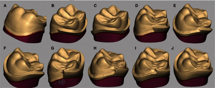
Fig. 1 Schematic representation of the shoulder variations in the zirconia core created in CAD software. The shoulder (1 mm in width) variations in core were incremental increases of 1 mm, 2 mm, and 3 mm in proximal and lingual (PL) height, and buccal (B) height respectively. A: no shoulder, B: PL 1mm, C: PL 1 mm and B 1 mm, D: PL 2 mm, E: PL 2 mm and B 1 mm, F: PL 2 mm and B 2 mm, G: PL 3 mm, H: PL 3 mm and B 1 mm, I: PL 3 mm and B 2 mm and J: PL 3 mm and B 3 mm.
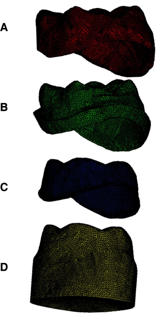
Fig. 2 CAD designed tooth/veneered zirconia crown system components. A: veneer porcelain, B: core, C: cement layers and D: tooth preparation.
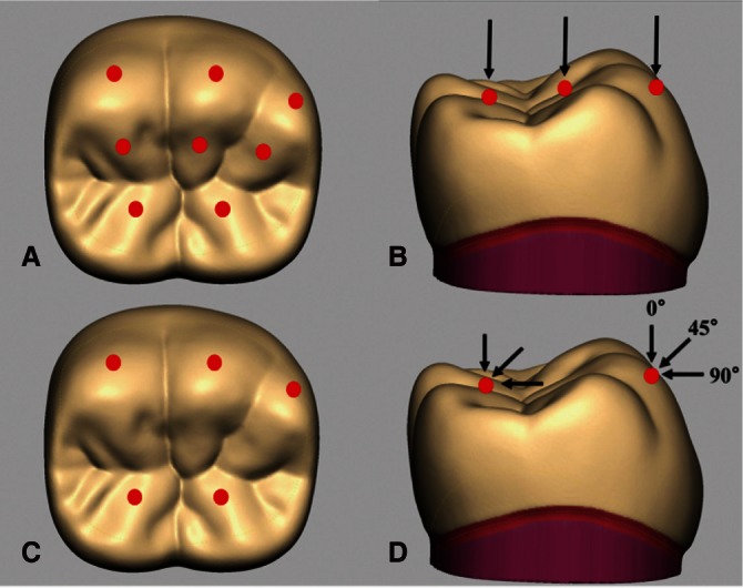
Fig. 3 Loading points and directions simulating maximum bite force (A and B) and masticatory force (C and D). A: Three points on the outer inclines of the buccal cusps, three points on the inner inclines of the buccal cusps, and two points on the inner inclines of the lingual cusps were loaded. B: A total load of 700 N was applied from the axial (vertical) direction. C: Three points on the outer inclines of the buccal cusps and two points on the inner inclines of the lingual cusps were loaded. D: A total load of 280 N was applied from three directions.
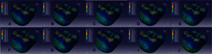
Fig. 4 Maximum principal stress distributions of 10 models subjected to maximum bite force. Maximum principal stress concentrated in the areas around loading points on the crown surface. A: Model 1, B: Model 2, C: Model 3, D: Model 4, E: Model 5, F: Model 6, G: Model 7, H: Model 8, I: Model 9 and J: Model 10.
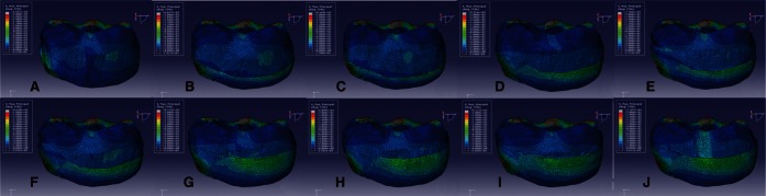
Fig. 5 Lingual side view of maximum principal stress distributions of 10 models subjected to maximum bite force. A: Model 1, B: Model 2, C: Model 3, D: Model 4, E: Model 5, F: Model 6, G: Model 7, H: Model 8, I: Model 9 and J: Model 10.
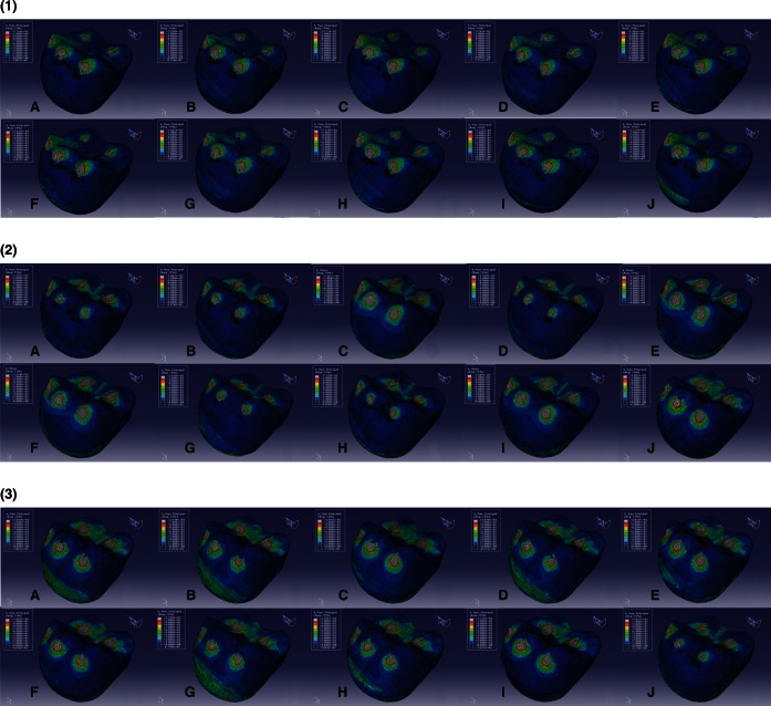
Fig. 6 Maximum principal stress distributions of 10 models subjected to masticatory force (under the application of loads from three directions). (1) load of 280 N at 0° to the to oth axis (vertical direction), (2) load of 280 N at 45° to the tooth axis, towards the lingual margin, and (3) load of 280 N at 90° to the tooth axis, towards the lingual surface (horizontal direction). A: Model 1, B: Model 2, C: Model 3, D: Model 4, E: Model 5, F: Model 6, G: Model 7, H: Model 8, I: Model 9 and J: Model 10.
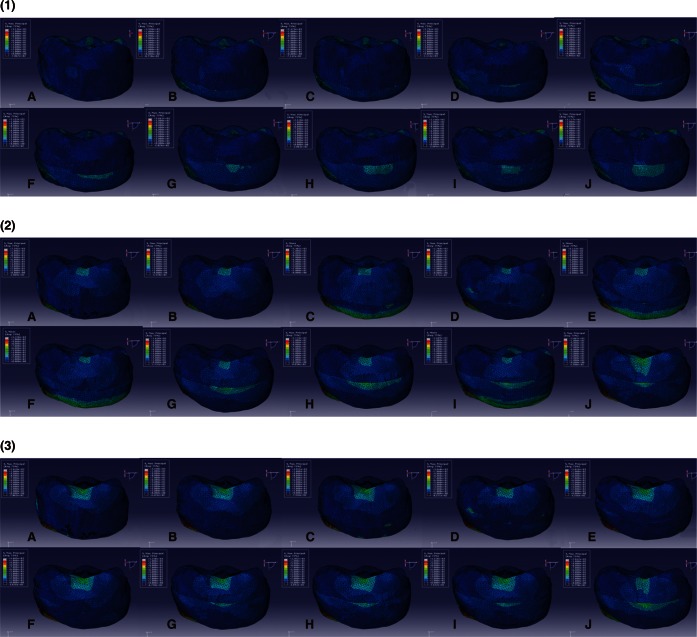
Fig. 7 Lingual side view of maximum principal stress distributions of 10 models subjected to masticatory force. (1) load of 280 N at 0° to the tooth axis (vertical direction), (2) load of 280 N at 45° to the tooth axis, towards the lingual margin, and (3) load of 280 N at 90° to the tooth axis, towards the lingual surface (horizontal direction). A: Model 1, B: Model 2, C: Model 3, B: Model 4, E: Model 5, F: Model 6, G: Model 7, H: Model 8, I: Model 9 and J: Model 10.
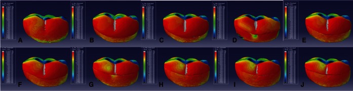
Fig. 8 Lingual side view of minimum principal stress distributions in the 10 models subjected to maximum bite force. A: Model 1, B: Model 2, C: Model 3, D: Model 4, E: Model 5, F: Model 6, G: Model 7, H: Model 8, I: Model 9 and J: Model 10.
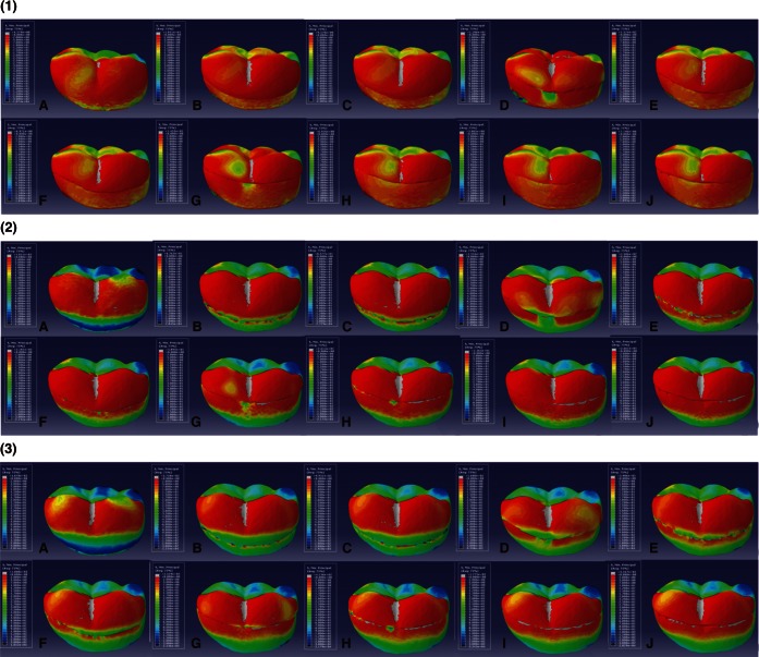
Fig. 9 Lingual side view of minimum principal stress distributions of 10 models subjected to masticatory force (under the application of loads from three directions). (1) load of 28 0 N at 0° to the tooth axis (vertical direction), (2) load of 280 N at 45° to the tooth axis, towards the lingual margin, and (3) load of 280 N at 90° to the tooth axis, towards the lingual surface (horizontal direction). A: Model 1, B: Model 2, C: Model 3, D: Model 4, E: Model 5, F: Model 6, G: Model 7, H: Model 8, I: Model 9 and J: Model 10.
Cited by 2 articles
-
A prospective clinical of lithium disilicate pressed zirconia and monolithic zirconia in posterior implant-supported prostheses: A 24-month follow-up
Kyoung-Woo Roh, Young-Chan Jeon, Chang-Mo Jeong, Mi-Jung Yun, Jung-Bo Huh, So-Hyoun Lee, Dong-Seok Yang, Eun-Bin Bae
J Korean Acad Prosthodont. 2019;57(2):134-141. doi: 10.4047/jkap.2019.57.2.134.Effect of polishing and glazing on the color and spectral distribution of monolithic zirconia
Hee-Kyung Kim, Sung-Hun Kim, Jai-Bong Lee, Jung-Suk Han, In-Sung Yeo
J Adv Prosthodont. 2013;5(3):296-304. doi: 10.4047/jap.2013.5.3.296.
Reference
-
1. Triwatana P, Nagaviroj N, Tulapornchai C. Clinical performance and failures of zirconia-based fixed partial dentures: a review literature. J Adv Prosthodont. 2012; 4:76–83. PMID: 22737311.
Article2. Imanishi A, Nakamura T, Ohyama T, Nakamura T. 3-D Finite element analysis of all-ceramic posterior crowns. J Oral Rehabil. 2003; 30:818–822. PMID: 12880406.
Article3. Fradeani M, Aquilano A. Clinical experience with Empress crowns. Int J Prosthodont. 1997; 10:241–247. PMID: 9484056.4. Fradeani M, D'Amelio M, Redemagni M, Corrado M. Five-year follow-up with Procera all-ceramic crowns. Quintessence Int. 2005; 36:105–113. PMID: 15732546.5. Fradeani M, Redemagni M. An 11-year clinical evaluation of leucite-reinforced glass-ceramic crowns: a retrospective study. Quintessence Int. 2002; 33:503–510. PMID: 12165985.6. Malament KA, Socransky SS. Survival of Dicor glass-ceramic dental restorations over 14 years. Part II: effect of thickness of Dicor material and design of tooth preparation. J Prosthet Dent. 1999; 81:662–667. PMID: 10347353.
Article7. Malament KA, Socransky SS. Survival of Dicor glass-ceramic dental restorations over 16 years. Part III: effect of luting agent and tooth or tooth-substitute core structure. J Prosthet Dent. 2001; 86:511–519. PMID: 11725279.
Article8. Sailer I, Fehér A, Filser F, Gauckler LJ, Lüthy H, Hämmerle CH. Five-year clinical results of zirconia frameworks for posterior fixed partial dentures. Int J Prosthodont. 2007; 20:383–388. PMID: 17695869.9. Sailer I, Fehér A, Filser F, Lüthy H, Gauckler LJ, Schärer P, Franz Hämmerle CH. Prospective clinical study of zirconia posterior fixed partial dentures: 3-year follow-up. Quintessence Int. 2006; 37:685–693. PMID: 17017630.10. Sailer I, Gottnerb J, Kanelb S, Hammerle CH. Randomized controlled clinical trial of zirconia-ceramic and metal-ceramic posterior fixed dental prostheses: a 3-year follow-up. Int J Prosthodont. 2009; 22:553–560. PMID: 19918588.11. Raigrodski AJ, Chiche GJ, Potiket N, Hochstedler JL, Mohamed SE, Billiot S, Mercante DE. The efficacy of posterior three-unit zirconium-oxide-based ceramic fixed partial dental prostheses: a prospective clinical pilot study. J Prosthet Dent. 2006; 96:237–244. PMID: 17052467.
Article12. Molin MK, Karlsson SL. Five-year clinical prospective evaluation of zirconia-based Denzir 3-unit FPDs. Int J Prosthodont. 2008; 21:223–227. PMID: 18548960.13. Roediger M, Gersdorff N, Huels A, Rinke S. Prospective evaluation of zirconia posterior fixed partial dentures: four-year clinical results. Int J Prosthodont. 2010; 23:141–148. PMID: 20305852.14. Schmitt J, Holst S, Wichmann M, Reich S, Gollner M, Hamel J. Zirconia posterior fixed partial dentures: a prospective clinical 3-year follow-up. Int J Prosthodont. 2009; 22:597–603. PMID: 19918596.15. Schmitter M, Mussotter K, Rammelsberg P, Stober T, Ohlmann B, Gabbert O. Clinical performance of extended zirconia frameworks for fixed dental prostheses: two-year results. J Oral Rehabil. 2009; 36:610–615. PMID: 19496928.
Article16. Vult von Steyern P, Carlson P, Nilner K. All-ceramic fixed partial dentures designed according to the DC-Zirkon technique. A 2-year clinical study. J Oral Rehabil. 2005; 32:180–187. PMID: 15707428.17. Tsumita M, Kokubo Y, Ohkubo C, Sakurai S, Fukushima S. Clinical evaluation of posterior all-ceramic FPDs (Cercon): a prospective clinical pilot study. J Prosthodont Res. 2010; 54:102–105. PMID: 20117970.
Article18. Wakabayashi N, Ona M, Suzuki T, Igarashi Y. Nonlinear finite element analyses: advances and challenges in dental applications. J Dent. 2008; 36:463–471. PMID: 18455859.
Article19. Eraslan O, Sevimay M, Usumez A, Eskitascioglu G. Effects of cantilever design and material on stress distribution in fixed partial dentures-a finite element analysis. J Oral Rehabil. 2005; 32:273–278. PMID: 15790382.20. Rafferty BT, Janal MN, Zavanelli RA, Silva NR, Rekow ED, Thompson VP, Coelho PG. Design features of a three-dimensional molar crown and related maximum principal stress. A finite element model study. Dent Mater. 2010; 26:156–163. PMID: 19857888.
Article21. Ausiello P, Apicella A, Davidson CL, Rengo S. 3D-finite element analyses of cusp movements in a human upper premolar, restored with adhesive resin-based composites. J Biomech. 2001; 34:1269–1277. PMID: 11522306.
Article22. Coelho PG, Silva NR, Thompson VP, Rekow D, Zhang G. Effect of proximal wall height on all-ceramic crown core stress distribution: a finite element analysis study. Int J Prosthodont. 2009; 22:78–86. PMID: 19260434.23. Ohlmann B, Marienburg K, Gabbert O, Hassel A, Gilde H, Rammelsberg P. Fracture-load values of all-ceramic cantilevered FPDs with different framework designs. Int J Prosthodont. 2009; 22:49–52. PMID: 19260427.24. Ulusoy M, Toksavul S. Fracture resistance of five different metal framework designs for metal-ceramic restorations. Int J Prosthodont. 2002; 15:571–574. PMID: 12475164.25. Rekow ED, Harsono M, Janal M, Thompson VP, Zhang G. Factorial analysis of variables influencing stress in all-ceramic crowns. Dent Mater. 2006; 22:125–132. PMID: 16000218.
Article26. Silva NR, Bonfante EA, Rafferty BT, Zavanelli RA, Rekow ED, Thompson VP, Coelho PG. Modified Y-TZP core design improves all-ceramic crown reliability. J Dent Res. 2011; 90:104–108. PMID: 21057036.
Article27. Lundgren D, Laurell L. Occlusal force pattern during chewing and biting in dentitions restored with fixed bridges of cross-arch extension. I. Bilateral end abutments. J Oral Rehabil. 1986; 13:57–71. PMID: 3511198.28. Gibbs CH, Mahan PE, Lundeen HC, Brehnan K, Walsh EK, Holbrook WB. Occlusal forces during chewing and swallowing as measured by sound transmission. J Prosthet Dent. 1981; 46:443–449. PMID: 6946215.
Article29. Magne P, Belser UC. Rationalization of shape and related stress distribution in posterior teeth: a finite element study using nonlinear contact analysis. Int J Periodontics Restorative Dent. 2002; 22:425–433. PMID: 12449302.
- Full Text Links
-
- Actions
-
Cited
- CITED
-
- Close
- Share
-
- Similar articles
-
- Biomechanical three-dimensional finite element analysis of monolithic zirconia crown with different cement type
- Finite element analysis of the influence of esthetic posts on incisors
- Influence of Crown Margin Design on the Stress Distribution in Maxillary Canine Restored by All-Ceramic Crown: A Finite Element Analysis
- Influence of various properties of post and core on the stress distribution in endodontically treated tooth
- An evaluation of the stress effect of different occlusion concepts on hybrid abutment and implant supported monolithic zirconia fixed prosthesis: A finite element analysis
