Focal epithelial hyperplasia arising after delivery of metal-ceramic fixed dental prosthesis
- Affiliations
-
- 1Department of Oral and Maxillofacial Surgery, Dental Reaserch Institute, Seoul National University, Seoul, Republic of Korea. smin5@snu.ac.kr
- 2Department of Oral and Maxillofacial Pathology, School of Dentistry, Kyung Hee University, Seoul, Republic of Korea.
- 3Department of Oral Pathology, College of Dentistry, Gangneung-Wonju National University, Gangneung, Republic of Korea.
- KMID: 2284731
- DOI: http://doi.org/10.4047/jap.2014.6.6.555
Abstract
- Focal epithelial hyperplasia (FEH) is a human papillomavirus (HPV)-induced alteration of the oral mucosa that presents with a clinically distinct appearance. While other HPV-infected lesions such as squamous papilloma, verruca vulgaris, and condyloma acuminatum involve the skin, oral mucosa, and genital mucosa, FEH occurs only in the oral mucosa. The affected oral mucosa exhibits multiple papules and nodules with each papule/nodule being flat-topped or sessile. The affected region resembles the normal color of oral mucosa rather than appearing as a white color since the epithelial surface is not hyperkeratinized. Almost all cases present with multiple sites of occurrence. This rare, benign epithelial proliferation is related to low-risk HPV, especially HPV-13 and -32, and is not transformed into carcinoma. We report a case of FEH that arose on the attached gingiva of an East Asian male adult related to prosthesis without detection of any HPV subtype in HPV DNA chip and sequencing.
Keyword
MeSH Terms
Figure
-
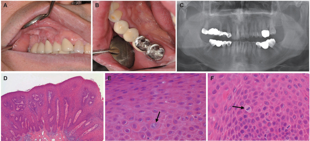
Fig. 1 Clinical intraoral view of the elevated nodules in the right upper posterior buccal gingiva (A) and in the left lower lingual attached gingiva (B) with a panoramic radiograph (C). A papillary proliferating pattern with acanthosis. Rete ridges were widened and elongated, but were not psoriasiform (original magnification, ×12.5) (D). Stratum spinosum layer showing a few mitosoid cells among the normal keratinocyte without brisk mitotic figures (original magnification, ×400) (E, F).
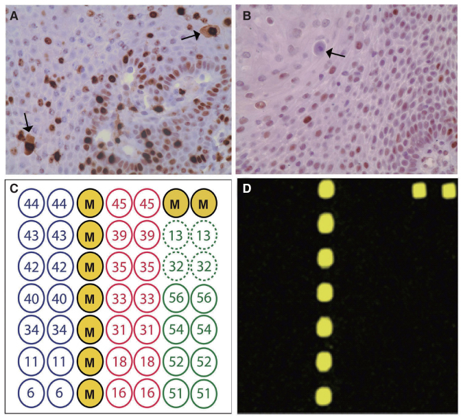
Fig. 2 Immunohistochemical results of Ki-67 and p53. Ki-67 was immunoreactive in the cells of the suprabasal layer as well as the basal cell layer, indicating hyperplasia of the keratinocytes (A), and p53-positive cells were scattered within the epithelium, but were few in number (B) (original magnification, ×400). Mitosoid cells were positive for Ki-67, but negative for p53. All the negative results of the DNA chip analysis (C, D).
Cited by 1 articles
-
Human papilloma virus in oral cancer
Soung Min Kim
J Korean Assoc Oral Maxillofac Surg. 2016;42(6):327-336. doi: 10.5125/jkaoms.2016.42.6.327.
Reference
-
1. Archard HO, Heck JW, Stanley HR. Focal epithelial hyperplasia: an unusual oral mucosal lesion found in indian children. Oral Surg Oral Med Oral Pathol. 1965; 20:201–212.2. Said AK, Leao JC, Fedele S, Porter SR. Focal epithelial hyperplasia - an update. J Oral Pathol Med. 2013; 42:435–442.3. Jayasooriya PR, Abeyratne S, Ranasinghe AW, Tilakaratne WM. Focal epithelial hyperplasia (Heck's disease): report of two cases with PCR detection of human papillomavirus DNA. Oral Dis. 2004; 10:240–243.4. Maschke J, Brauns TC, Goos M. Imiquimod for the topical treatment of focal epithelial hyperplasia (Heck disease) in a child. J Dtsch Dermatol Ges. 2004; 2:848–850.5. Kim SM, Myoung H, Choung PH, Kim MJ, Lee SK, Lee JH. Metastatic leiomyosarcoma in the oral cavity: case report with protein expression profiles. J Craniomaxillofac Surg. 2009; 37:454–460.6. Bennett LK, Hinshaw M. Heck's disease: diagnosis and susceptibility. Pediatr Dermatol. 2009; 26:87–89.7. AlSheddi MA, Faden AA. Multifocal epithelial hyperplasia in an adult female. Hong Kong J Dermatol Venereol. 2012; 20:23–27.8. Padayachee A, van Wyk CW. Human papillomavirus (HPV) DNA in focal epithelial hyperplasia by in situ hybridization. J Oral Pathol Med. 1991; 20:210–214.9. Ledesma-Montes C, Vega-Memije E, Garcés-Ortíz M, Cardiel-Nieves M, Juárez-Luna C. Multifocal epithelial hyperplasia. Report of nine cases. Med Oral Patol Oral Cir Bucal. 2005; 10:394–401.10. Pfister H, Hettich I, Runne U, Gissmann L, Chilf GN. Characterization of human papillomavirus type 13 from focal epithelial hyperplasia Heck lesions. J Virol. 1983; 47:363–366.11. Beaudenon S, Praetorius F, Kremsdorf D, Lutzner M, Worsaae N, Pehau-Arnaudet G, Orth G. A new type of human papillomavirus associated with oral focal epithelial hyperplasia. J Invest Dermatol. 1987; 88:130–135.12. Burd EM. Human papillomavirus and cervical cancer. Clin Microbiol Rev. 2003; 16:1–17.13. Falaki F, Amir Chaghmaghi M, Pakfetrat A, Delavarian Z, Mozaffari PM, Pazooki N. Detection of human papilloma virus DNA in seven cases of focal epithelial hyperplasia in Iran. J Oral Pathol Med. 2009; 38:773–776.14. Ozden B, Gunduz K, Gunhan O, Ozden FO. A Case Report of Focal Epithelial Hyperplasia (Heck's disease) with PCR Detection of Human Papillomavirus. J Maxillofac Oral Surg. 2011; 10:357–360.15. Choi YD, Jung WW, Nam JH, Choi HS, Park CS. Detection of HPV genotypes in cervical lesions by the HPV DNA Chip and sequencing. Gynecol Oncol. 2005; 98:369–375.16. Ledesma-Montes C, Garces-ortiz M, Edmundo B. Multifocal epithelial hyperplasia. An unusual lesion. Webmedcentral Pathology. 2012; 3:WMC003003.17. Ciszewski A, Baraniak M, Urbanek-Brychczyńska M. Corrosion by galvanic coupling between amalgam and different chromium-based alloys. Dent Mater. 2007; 23:1256–1261.18. Padbury A Jr, Eber R, Wang HL. Interactions between the gingiva and the margin of restorations. J Clin Periodontol. 2003; 30:379–385.
- Full Text Links
-
- Actions
-
Cited
- CITED
-
- Close
- Share
-
- Similar articles
-
- Fabrication of a metal-ceramic crown to fit an existing partial removable dental prosthesis using ceramic pressed to metal technique: a clinical report
- Clinical cases of implant-supported fixed dental prosthesis using modified lingual screw system (T-screw system)
- Evaluation of the marginal and internal gap of metal-ceramic crown fabricated with a selective laser sintering technology: two- and three-dimensional replica techniques
- The effet of cooling rate on the residual stresses in the veneer ceramics of zirconia-ceramic restorations: a literature review
- An alternative method to convert fractured metal ceramic surveyed crown into a complete contour zirconia surveyed crown using CAD-CAM technology under anticancer treatments: a clinical report
