A prospective study on the effectiveness of newly developed autogenous tooth bone graft material for sinus bone graft procedure
- Affiliations
-
- 1Department of Oral and Maxillofacial Surgery, Section of Dentistry, Korea University Anam Hospital, Seoul, Republic of Korea.
- 2Department of Dental Biomaterials Science and Dental Research Institute, School of Dentistry, Seoul National University, Seoul, Republic of Korea.
- 3Department of Dentistry & Dental Research Institute, School of Dentistry, Seoul National University, Seoul, Republic of Korea. kyk0505@snubh.org
- 4Department of Oral Pathology and Dental Research Institute, School of Dentistry, Seoul National University, Seoul, Republic of Korea.
- 5Department of Oral and Maxillofacial Surgery, Section of Dentistry, Seoul National University Bundang Hospital, Seongnam, Republic of Korea.
- KMID: 2284728
- DOI: http://doi.org/10.4047/jap.2014.6.6.528
Abstract
- PURPOSE
The purpose of this prospective study was to evaluate the effectiveness of newly developed autogenous tooth bone graft material (AutoBT)application for sinus bone graft procedure.
MATERIALS AND METHODS
The patients with less than 5.0 mm of residual bone height in maxillary posterior area were enrolled. For the sinus bone graft procedure, Bio-Oss was grafted in control group and AutoBT powder was grafted in experimental group. Clinical and radiographic examination were done for the comparison of grafted materials in sinus cavity between groups. At 4 months after sinus bone graft procedure, biopsy specimens were analyzed by microcomputed tomography and histomorphometric examination for the evaluation of healing state of bone graft site.
RESULTS
In CT evaluation, there was no difference in bone density, bone height and sinus membrane thickness between groups. In microCT analysis, there was no difference in total bone volume, new bone volume, bone mineral density of new bone between groups. There was significant difference trabecular thickness (0.07 microm in Bio-Oss group Vs. 0.08 microm in AutoBT group) (P=.006). In histomorphometric analysis, there was no difference in new bone formation, residual graft material, bone marrow space between groups. There was significant difference osteoid thickness (8.35 microm in Bio-Oss group Vs. 13.12 microm in AutoBT group) (P=.025).
CONCLUSION
AutoBT could be considered a viable alternative to the autogenous bone or other bone graft materials in sinus bone graft procedure.
MeSH Terms
Figure
-
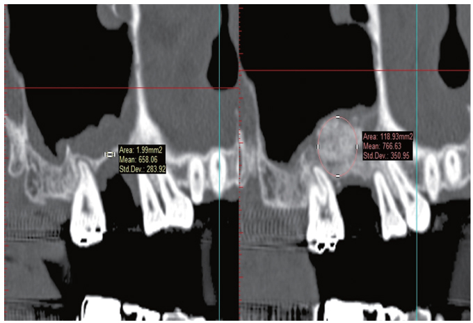
Fig. 1 Measurement of bone density by Simplant software.
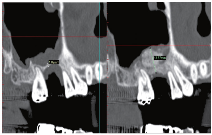
Fig. 2 Measurement of bone height by Simplant software.
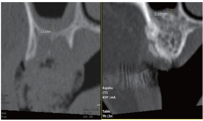
Fig. 3 Measurement of sinus membrane thickness by INFINITT PACS software.
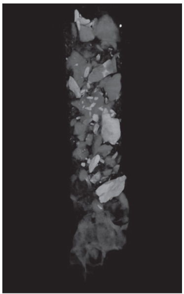
Fig. 4 MicroCT image.
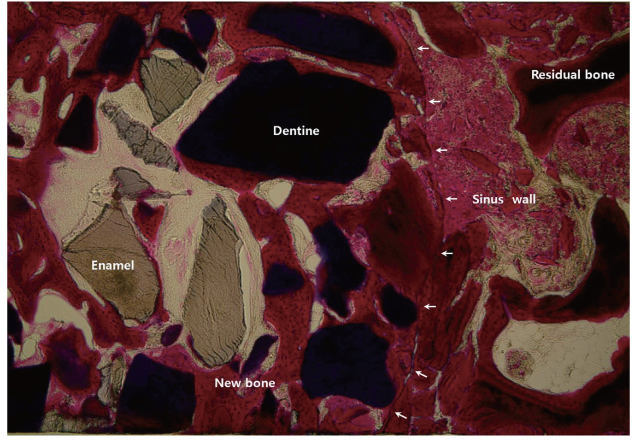
Fig. 5 Matured lamellar bone showing bony integration between residual bone and maxillary sinus was detected. Bone formation was found surrounding both enamel and dentin portion of AutoBT material. New bone bridges between graft materials were also detected (Masson's trichrome staining, ×35).
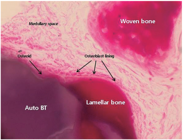
Fig. 6 Newly-formed and matured lamellar bone was detected surrounding AutoBT material, osteoblasts covering newly-formed bone accumulated osteoid. Active woven bone formation was also detected. Medullary space formation composed of well vascularized connective tissue was also detected within new bone (Masson's trichrome staining, ×400).
Cited by 2 articles
-
Comparison of immunohistochemical analysis on sinus augmentation using demineralized tooth graft and bovine bone
Dong-Seok Sohn, Ji-Rak Kim, Hyung-Gyun Kim, Hyun-Suk Choi, Yong-Suk Moon
J Korean Assoc Oral Maxillofac Surg. 2021;47(4):269-278. doi: 10.5125/jkaoms.2021.47.4.269.Autogenous tooth bone graft block for sinus augmentation with simultaneous implant installation: a technical note
Kwang-Ho Lee, Young-Kyun Kim, Woo-Jin Cho, In-Woong Um, Masaru Murata, Masaharu Mitsugi
J Korean Assoc Oral Maxillofac Surg. 2015;41(5):284-289. doi: 10.5125/jkaoms.2015.41.5.284.
Reference
-
1. Berglundh T, Lindhe J. Healing around implants placed in bone defects treated with Bio-Oss. An experimental study in the dog. Clin Oral Implants Res. 1997; 8:117–124.2. Piattelli M, Favero GA, Scarano A, Orsini G, Piattelli A. Bone reactions to anorganic bovine bone (Bio-Oss) used in sinus augmentation procedures: a histologic long-term report of 20 cases in humans. Int J Oral Maxillofac Implants. 1999; 14:835–840.3. Carmagnola D, Adriaens P, Berglundh T. Healing of human extraction sockets filled with Bio-Oss. Clin Oral Implants Res. 2003; 14:137–143.4. Caubet J, Petzold C, Sáez-Torres C, Morey M, Iriarte JI, Sánchez J, Torres JJ, Ramis JM, Monjo M. Sinus graft with safescraper: 5-year results. J Oral Maxillofac Surg. 2011; 69:482–490.5. Kim YK, Kim SG, Byeon JH, Lee HJ, Um IU, Lim SC, Kim SY. Development of a novel bone grafting material using autogenous teeth. Oral Surg Oral Med Oral Pathol Oral Radiol Endod. 2010; 109:496–503.6. Kim YK, Kim SG, Oh JS, Jin SC, Son JS, Kim SY, Lim SY. Analysis of the inorganic component of autogenous tooth bone graft material. J Nanosci Nanotechnol. 2011; 11:7442–7445.7. Kim YK, Lee JK, Kim KW, Um IW, Murata M. Healing Mechanism and Clinical Application of Autogenous Tooth Bone Graft Material. In : Pignatello R, editor. Advances in Biomaterials Science and Biomedical Applications [Internet]. Rijeka, Croatia: InTech;2013. p. 405–435.8. Kim YK, Yi YJ. Horizontal ridge augmentation using ridge expansion and autogenous tooth bone graft: a case report. J Dent Rehabil Appl Sci. 2011; 27:109–115.9. Kim YK, Kim SG, Um IW. Vertical and horizontal ridge augmentation using autogenous tooth bone graft materials: case report. J Korean Assoc Maxillofac Plast Reconstr Surg. 2011; 33:166–170.10. Kim YK, Kim SG, Kim KW, Um IW. Extraction socket preservation and reconstruction using autogenous tooth bone graft: Case report. J Korean Assoc Maxillofac Plast Reconstr Surg. 2011; 33:264–269.11. Kim YK, Choi YH. Tooth autotransplantation with autogenous tooth- bone graft: a case report. J Korean Dent Sci. 2011; 4:79–84.12. Lee JY, Kim YK, Kim SG, Lim SC. Histomorphometric study of sinus bone graft using various graft material. J Dent Rehab App Sci. 2011; 27:141–147.13. Kim YK, Lee HJ, Kim KW, Kim SG, Um IW. Guide bone regeneration using autogenous teeth: case reports. J Korean Assoc Oral Maxillofac Surg. 2011; 37:142–147.14. Jeong KI, Kim SG, Kim YK, Oh JS, Jeong MA, Park JJ. Clinical study of graft materials using autogenous teeth in maxillary sinus augmentation. Implant Dent. 2011; 20:471–475.15. Lee JY, Kim YK. Retrospective cohort study of autogenous tooth bone graft. Oral Biol Res. 2012; 36:39–43.16. Kim YK, Um IW, Murata M. Tooth bank system for bone regeneration-Safety report-. J Hard Tissue Biol. 2014; 23:371–376.17. Jensen OT. The sinus bone graft. 2nd ed. Chicago: Quintessence Publ;2006. p. 103–125.18. Kim YK, Kim SG, Park JY, Yi YJ, Bae JH. Comparison of clinical outcomes of sinus bone graft with simultaneous implant placement: 4-month and 6-month final prosthetic loading. Oral Surg Oral Med Oral Pathol Oral Radiol Endod. 2011; 111:164–169.19. Janner SF, Caversaccio MD, Dubach P, Sendi P, Buser D, Bornstein MM. Characteristics and dimensions of the Schneiderian membrane: a radiographic analysis using cone beam computed tomography in patients referred for dental implant surgery in the posterior maxilla. Clin Oral Implants Res. 2011; 22:1446–1453.20. Phothikhun S, Suphanantachat S, Chuenchompoonut V, Nisapakultorn K. Cone-beam computed tomographic evidence of the association between periodontal bone loss and mucosal thickening of the maxillary sinus. J Periodontol. 2012; 83:557–564.21. Tajima N, Ohba S, Sawase T, Asahina I. Evaluation of sinus floor augmentation with simultaneous implant placement using platelet-rich fibrin as sole grafting material. Int J Oral Maxillofac Implants. 2013; 28:77–83.22. Nevins M, Kirker-Head C, Nevins M, Wozney JA, Palmer R, Graham D. Bone formation in the goat maxillary sinus induced by absorbable collagen sponge implants impregnated with recombinant human bone morphogenetic protein-2. Int J Periodontics Restorative Dent. 1996; 16:8–19.23. Kühl S, Götz H, Brochhausen C, Jakse N, Filippi A, d'Hoedt B, Kreisler M. The influence of substitute materials on bone density after maxillary sinus augmentation: a microcomputed tomography study. Int J Oral Maxillofac Implants. 2012; 27:1541–1546.24. Huang HL, Chen MY, Hsu JT, Li YF, Chang CH, Chen KT. Three-dimensional bone structure and bone mineral density evaluations of autogenous bone graft after sinus augmentation: a microcomputed tomography analysis. Clin Oral Implants Res. 2012; 23:1098–1103.25. Lundgren S, Moy P, Johansson C, Nilsson H. Augmentation of the maxillary sinus floor with particulated mandible: a histologic and histomorphometric study. Int J Oral Maxillofac Implants. 1996; 11:760–766.26. Chackartchi T, Iezzi G, Goldstein M, Klinger A, Soskolne A, Piattelli A, Shapira L. Sinus floor augmentation using large (1-2 mm) or small (0.25-1 mm) bovine bone mineral particles: a prospective, intra-individual controlled clinical, microcomputerized tomography and histomorphometric study. Clin Oral Implants Res. 2011; 22:473–480.27. Kim DM, Nevins ML, Camelo M, Camelo JM, Schupbach P, Hanratty JJ, Uzel NG, Nevins M. The efficacy of demineralized bone matrix and cancellous bone chips for maxillary sinus augmentation. Int J Periodontics Restorative Dent. 2009; 29:415–423.28. Gapski R, Neiva R, Oh TJ, Wang HL. Histologic analyses of human mineralized bone grafting material in sinus elevation procedures: a case series. Int J Periodontics Restorative Dent. 2006; 26:59–69.29. Cammack GV, Nevins M, Clem DS, Hatch JP, Mellonig JT. Histologic evaluation of mineralized and demineralized freeze-dried bone allograft for ridge and sinus augmentations. Int J Periodontics Restorative Dent. 2005; 25:231–237.30. Chaushu G, Vered M, Mardinger O, Nissan J. Histomorphometric analysis after maxillary sinus floor augmentation using cancellous bone-block allograft. J Periodontol. 2010; 81:1147–1152.31. Scarano A, Degidi M, Iezzi G, Pecora G, Piattelli M, Orsini G, Caputi S, Perrotti V, Mangano C, Piattelli A. Maxillary sinus augmentation with different biomaterials: a comparative histologic and histomorphometric study in man. Implant Dent. 2006; 15:197–207.32. Szabó G, Huys L, Coulthard P, Maiorana C, Garagiola U, Barabás J, Németh Z, Hrabák K, Suba Z. A prospective multicenter randomized clinical trial of autogenous bone versus beta-tricalcium phosphate graft alone for bilateral sinus elevation: histologic and histomorphometric evaluation. Int J Oral Maxillofac Implants. 2005; 20:371–381.33. Kim YK, Yun PY, Lim SC, Kim SG, Lee HJ, Ong JL. Clinical evaluations of OSTEON as a new alloplastic material in sinus bone grafting and its effect on bone healing. J Biomed Mater Res B Appl Biomater. 2008; 86:270–277.34. Kolerman R, Goshen G, Joseph N, Kozlovsky A, Shetty S, Tal H. Histomorphometric analysis of maxillary sinus augmentation using an alloplast bone substitute. J Oral Maxillofac Surg. 2012; 70:1835–1843.35. Kurkcu M, Benlidayi ME, Cam B, Sertdemir Y. Anorganic bovine-derived hydroxyapatite vs β-tricalcium phosphate in sinus augmentation: a comparative histomorphometric study. J Oral Implantol. 2012; 38:519–526.36. Ozyuvaci H, Bilgiç B, Firatli E. Radiologic and histomorphometric evaluation of maxillary sinus grafting with alloplastic graft materials. J Periodontol. 2003; 74:909–915.37. Karabuda C, Ozdemir O, Tosun T, Anil A, Olgaç V. Histological and clinical evaluation of 3 different grafting materials for sinus lifting procedure based on 8 cases. J Periodontol. 2001; 72:1436–1442.38. Wallace SS, Froum SJ, Cho SC, Elian N, Monteiro D, Kim BS, Tarnow DP. Sinus augmentation utilizing anorganic bovine bone (Bio-Oss) with absorbable and nonabsorbable membranes placed over the lateral window: histomorphometric and clinical analyses. Int J Periodontics Restorative Dent. 2005; 25:551–559.39. Zhang Y, Tangl S, Huber CD, Lin Y, Qiu L, Rausch-Fan X. Effects of Choukroun's platelet-rich fibrin on bone regeneration in combination with deproteinized bovine bone mineral in maxillary sinus augmentation: a histological and histomorphometric study. J Craniomaxillofac Surg. 2012; 40:321–328.40. Orsini G, Scarano A, Piattelli M, Piccirilli M, Caputi S, Piattelli A. Histologic and ultrastructural analysis of regenerated bone in maxillary sinus augmentation using a porcine bone-derived biomaterial. J Periodontol. 2006; 77:1984–1990.41. Galindo-Moreno P, Avila G, Fernández-Barbero JE, Aguilar M, Sánchez-Fernández E, Cutando A, Wang HL. Evaluation of sinus floor elevation using a composite bone graft mixture. Clin Oral Implants Res. 2007; 18:376–382.42. Peleg M, Garg AK, Misch CM, Mazor Z. Maxillary sinus and ridge augmentations using a surface-derived autogenous bone graft. J Oral Maxillofac Surg. 2004; 62:1535–1544.43. Artzi Z, Kozlovsky A, Nemcovsky CE, Weinreb M. The amount of newly formed bone in sinus grafting procedures depends on tissue depth as well as the type and residual amount of the grafted material. J Clin Periodontol. 2005; 32:193–199.44. Hanisch O, Lozada JL, Holmes RE, Calhoun CJ, Kan JY, Spiekermann H. Maxillary sinus augmentation prior to placement of endosseous implants: A histomorphometric analysis. Int J Oral Maxillofac Implants. 1999; 14:329–336.45. Gonshor A, McAllister BS, Wallace SS, Prasad H. Histologic and histomorphometric evaluation of an allograft stem cell-based matrix sinus augmentation procedure. Int J Oral Maxillofac Implants. 2011; 26:123–131.46. Tarnow DP, Wallace SS, Froum SJ, Rohrer MD, Cho SC. Histologic and clinical comparison of bilateral sinus floor elevations with and without barrier membrane placement in 12 patients: Part 3 of an ongoing prospective study. Int J Periodontics Restorative Dent. 2000; 20:117–125.47. Froum SJ, Tarnow DP, Wallace SS, Rohrer MD, Cho SC. Sinus floor elevation using anorganic bovine bone matrix (OsteoGraf/N) with and without autogenous bone: a clinical, histologic, radiographic, and histomorphometric analysis--Part 2 of an ongoing prospective study. Int J Periodontics Restorative Dent. 1998; 18:528–543.48. Lambert F, Léonard A, Drion P, Sourice S, Layrolle P, Rompen E. Influence of space-filling materials in subantral bone augmentation: blood clot vs. autogenous bone chips vs. bovine hydroxyapatite. Clin Oral Implants Res. 2011; 22:538–545.49. Lee YM, Shin SY, Kim JY, Kye SB, Ku Y, Rhyu IC. Bone reaction to bovine hydroxyapatite for maxillary sinus floor augmentation: histologic results in humans. Int J Periodontics Restorative Dent. 2006; 26:471–481.50. De Leonardis D, Pecora GE. Prospective study on the augmentation of the maxillary sinus with calcium sulfate: histological results. J Periodontol. 2000; 71:940–947.
- Full Text Links
-
- Actions
-
Cited
- CITED
-
- Close
- Share
-
- Similar articles
-
- Familial tooth bone graft for ridge and sinus augmentation: a report of two cases
- Bone graft material using teeth
- Evaluation of the Healing Process of Autogenous Tooth Bone Graft Material Nine Months after Sinus Bone Graft: Micromorphometric and Histological Evaluation
- Clinical Study on the Efficacy of the Autogenous Tooth Bone Graft Material (AutoBT)
- Clinical Study on the Alveolar Bone Repair Capacity of Dentin Matrix Block
