J Adv Prosthodont.
2015 Jun;7(3):199-206. 10.4047/jap.2015.7.3.199.
Impact of surface roughness of gypsum materials on adaptation of zirconia cores
- Affiliations
-
- 1Institute for Health Science, College of Health Science, Korea University, Seoul, Republic of Korea. noreason07@korea.ac.kr
- 2Department of Dental Technology, School of Medical and Public Health, Kyungdong University, Goseong, Gangwondo, Republic of Korea.
- KMID: 2284705
- DOI: http://doi.org/10.4047/jap.2015.7.3.199
Abstract
- PURPOSE
The present study investigated the influences of various gypsum materials on the precision of fit of CAD/CAM-fabricated prostheses and analyzed their correlation with surface roughness.
MATERIALS AND METHODS
The master model of the mandibular right first molar was replicated, and four experimental groups based on two types of Type IV stone (GC Fujirock EP, Die keen) and two types of scannable stone (Aesthetic-Basegold, Everest Rock) were created to include a total of 40 specimens, 10 in each group. The surface roughness of the working models for the respective experimental groups was measured. Once the zirconia cores had been fabricated, the marginal and internal fits were measured with a digital microscope using the silicone replica technique. The mean and standard deviation of the respective points of measurement were computed and analyzed through the one-way ANOVA and Tukey's HSD test. The correlation between surface roughness and the precision of fit of the zirconia core was analyzed using the Pearson correlation analysis (alpha=.05).
RESULTS
The zirconia cores fabricated from the scannable stone working models exhibited a superior precision of fit as compared to those fabricated from the Type IV stone working models. The correlation analysis results showed a clear positive correlation between surface roughness and the precision of fit of zirconia cores in all of the experimental groups (P<.05).
CONCLUSION
The results confirmed that the surface roughness of dental working models has a decisive influence on the precision of fit of zirconia cores.
MeSH Terms
Figure
-

Fig. 1 Master model.
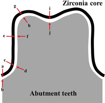
Fig. 2 Zirconia core to abutment teeth diagram showing measurement points to determine adaptation.

Fig. 3 Photograph of cross section of silicone replica measured at ×160 magnification using a digital microscope.

Fig. 4 Multifocus images of the surfaces of various gypsum materials (×20). (A) TY1, (B) TY2, (C) SCAN1, and (D) SCAN2 groups.
Reference
-
1. Al-Abidi K, Ellakwa A. The effect of adding a stone base on the accuracy of working casts using different types of dental stone. J Contemp Dent Pract. 2006; 7:17–28.2. Anusavice KJ. Phillip's science of dental material. 11th ed. Philadelphia: WB Saunders;2003. p. 621–654.3. ISO 6873. Dental gypsum products. International Organization for Standardization. 3rd ed. Geneva: Switzerland;2013.4. Retrieved April 12, 2014. Available from http://en.zhermack.com/ImagePub.aspx?id=120081.pdf.5. Kim KB, Kim JH, Kim WC, Kim HY, Kim JH. Evaluation of the marginal and internal gap of metal-ceramic crown fabricated with a selective laser sintering technology: two- and three-dimensional replica techniques. J Adv Prosthodont. 2013; 5:179–186.6. American Dental Association. Specification No. 25 for dental gypsum products. Certification program for dental materials. Chicago: American Dental Association;1990. p. 1–14.7. Millstein PL. Determining the accuracy of gypsum casts made from type IV dental stone. J Oral Rehabil. 1992; 19:239–243.8. Duke P, Moore BK, Haug SP, Andres CJ. Study of the physical properties of type IV gypsum, resin-containing, and epoxy die materials. J Prosthet Dent. 2000; 83:466–473.9. Kim JH, Kim KB, Kim WC, Kim JH, Kim HY. Accuracy and precision of polyurethane dental arch models fabricated using a three-dimensional subtractive rapid prototyping method with an intraoral scanning technique. Korean J Orthod. 2014; 44:69–76.10. Linke BA, Nicholls JI, Faucher RR. Distortion analysis of stone casts made from impression materials. J Prosthet Dent. 1985; 54:794–802.11. DeLong R, Pintado MR, Ko CC, Hodges JS, Douglas WH. Factors influencing optical 3D scanning of vinyl polysiloxane impression materials. J Prosthodont. 2001; 10:78–85.12. Rodriguez JM, Curtis RV, Bartlett DW. Surface roughness of impression materials and dental stones scanned by non-contacting laser profilometry. Dent Mater. 2009; 25:500–505.13. Beuer F, Aggstaller H, Edelhoff D, Gernet W, Sorensen J. Marginal and internal fits of fixed dental prostheses zirconia retainers. Dent Mater. 2009; 25:94–102.14. Kokubo Y, Nagayama Y, Tsumita M, Ohkubo C, Fukushima S, Vult von Steyern P. Clinical marginal and internal gaps of In-Ceram crowns fabricated using the GN-I system. J Oral Rehabil. 2005; 32:753–758.15. Williams GJ, Wild S, Bates JF. A study of some factors affecting the surface properties of dental stone. Br Dent J. 1984; 156:46–53.16. Keuter FM, Davidson CL. Surface roughness of dental stone casts from alginate impressions. J Dent. 1986; 14:23–28.17. Nanami T, Ostlund SG. Surface texture of stone model setting against elastomer impression materials. Swed Dent J. 1984; 8:251–255.18. Ender A, Mehl A. Influence of scanning strategies on the accuracy of digital intraoral scanning systems. Int J Comput Dent. 2013; 16:11–21.19. Karl M, Graef F, Schubinski P, Taylor T. Effect of intraoral scanning on the passivity of fit of implant-supported fixed dental prostheses. Quintessence Int. 2012; 43:555–562.20. Luthardt RG, Sandkuhl O, Herold V, Walter MH. Accuracy of mechanical digitizing with a CAD/CAM system for fixed restorations. Int J Prosthodont. 2001; 14:146–151.21. Reports of councils and bureaus: Revised American National Standards Institute/American Dental Association Specification No. 8 for zinc phosphate cement. J Am Dent Assoc. 1978; 96:121–123.22. Sorensen SE, Larsen IB, Jörgensen KD. Gingival and alveolar bone reaction to marginal fit of subgingival crown margins. Scand J Dent Res. 1986; 94:109–114.23. McLean JW, von Fraunhofer JA. The estimation of cement film thickness by an in vivo technique. Br Dent J. 1971; 131:107–111.24. Jorgensen KD, Esbensen AL. The relationship between the film thickness of zinc phosphate cement and the retention of veneer crowns. Acta Odontol Scand. 1968; 26:169–175.25. Passon C, Lambert RH, Lambert RL, Newman S. The effect of multiple layers of die-spacer on crown retention. Oper Dent. 1992; 17:42–49.26. Persson AS, Andersson M, Odén A, Sandborgh-Englund G. Computer aided analysis of digitized dental stone replicas by dental CAD/CAM technology. Dent Mater. 2008; 24:1123–1130.
- Full Text Links
-
- Actions
-
Cited
- CITED
-
- Close
- Share
-
- Similar articles
-
- Comparison of surface topography and roughness in different yttrium oxide compositions of dental zirconia after grinding and polishing
- Comparative study in marginal adaptation of zirconia cores fabricated with 3 different CAD/CAM systems
- Investigation of effect of zirconia on osseointegration by surface treatments
- The effect of various polishing systems on surface roughness and phase transformation of monolithic zirconia
- The Effect of ZrO2 Slurry Application to the Pre-sintered Zirconia Surface on Bonding Strength
