A case of double primary cancers of uterine endometrium and bladder with unusual histology
- Affiliations
-
- 1Department of Obstetrics and Gynecology, St. Vincent's Hospital, The Catholic University of Korea, Suwon, Korea. dcpark@catholic.ac.kr
- KMID: 2274081
- DOI: http://doi.org/10.5468/KJOG.2011.54.9.543
Abstract
- A case of a moderate differentiated endometrial carcinoma of the uterus with a synchronous poorly differentiated bladder cancer is reported. A review of the literature revealed that simultaneous presentation of primary endometrial and bladder neoplasm is rare and usually related to low-stage bladder lesions in contrast to our case with undifferentiated and the deep myometrial invasion of bladder lesion. A 79-year-old woman with endometrial cancer stage IB was performed of total abdominal hysterectomy with bilateral salpingo-oophorectomy, pelvic and para-aortic lymph nodes dissection and adjuvant concurrent cisplatin-radiation therapy. After treatment, she complained intermittent gross hematuria. She was performed the bladder mucosal biopsy and finally diagnoses with poorly differentiated carcinoma of bladder. She received transurethral resection of bladder tumor alone without total cystectomy or any other adjuvant treatment due to her refusal. Her condition is tolerable except intermittent hematuria and anemia.
MeSH Terms
Figure
-
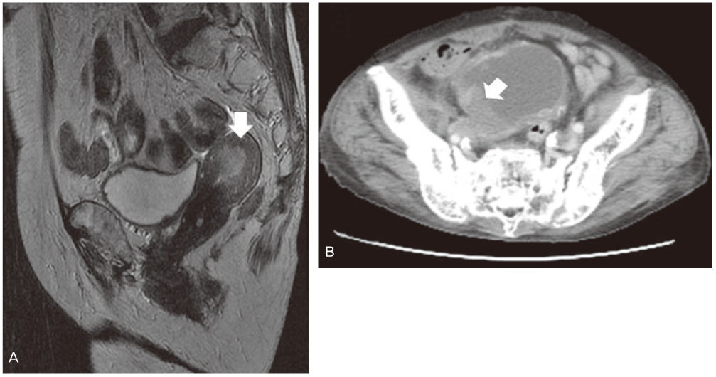
Fig. 1 (A) Pelvic magnetic resonance imaging. About 2.8×2.6×2.4 cm sized T2 intermediate signal intensity mass (arrow) within the endometrial cavity and thinning of myometrium, especially right side body and fundus. (B) Pelvic computed tomography. Irregular lobulated tumor (arrow) around right posterior inferior wall of the urinary bladder with suspicious adjacent perivascular retroperitoneal space infiltration along the right internal iliac vessels.
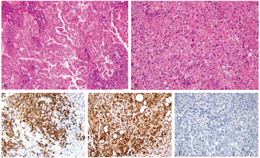
Fig. 2 In the uterus, typical endometrioid adenocarcinoma invading myometrium is noted (A). The lesion in the urinary bladder is morphologically different from that of the uterus, and composed of highly pleomorphic malignant cells without forming glandular structure (B). Immunohistologically, these tumor cells show positive reaction to cytokeratin (CK)-7 (C) and vimentin (D), but negative reaction to CK20 (E). So, for the lesion in the urinary bladder, poorly differentiated carcinoma or carcinosarcoma is suspected. Magnification, ×200.
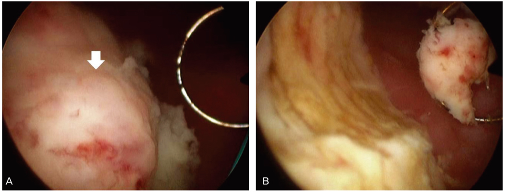
Fig. 3 Cystoscopic finding of transurethral resection of bladder tumor. Arrow indicated protruding tumor mass.
Reference
-
1. Sheu BC, Lin HH, Chen CK, Chao KH, Shun CT, Huang SC. Synchronous primary carcinomas of the endometrium and ovary. Int J Gynaecol Obstet. 1995. 51:141–146.2. Eisner RF, Nieberg RK, Berek JS. Synchronous primary neoplasms of the female reproductive tract. Gynecol Oncol. 1989. 33:335–339.3. Castro IM, Connell PP, Waggoner S, Rotmensch J, Mundt AJ. Synchronous ovarian and endometrial malignancies. Am J Clin Oncol. 2000. 23:521–525.4. Prat J, Matias-Guiu X, Barreto J. Simultaneous carcinoma involving the endometrium and the ovary. A clinicopathologic, immunohistochemical, and DNA flow cytometric study of 18 cases. Cancer. 1991. 68:2455–2459.5. Prat J. Clonality analysis in synchronous tumors of the female genital tract. Hum Pathol. 2002. 33:383–385.6. Lynch HT, Harris RE, Lynch PM, Guirgis HA, Lynch JF, Bardawil WA. Role of heredity in multiple primary cancer. Cancer. 1977. 40:1849–1854.7. Ayhan A, Yalcin OT, Tuncer ZS, Gürgan T, Küçükali T. Synchronous primary malignancies of the female genital tract. Eur J Obstet Gynecol Reprod Biol. 1992. 45:63–66.8. Schröcksnadel H, Fuith LC, Hetzel H. Multiple cancers in gynecologic oncology. Geburtshilfe Frauenheilkd. 1988. 48:710–714.9. Chen F, Shen K, Lang JH, Huang HF, Wu M. Clinical features and prognostic of double primary carcinoma of uterine corpus and the ovary. Zhonghua Yi Xue Za Zhi. 2005. 85:1257–1260.10. Papathanasiou K, Tolikas A, Dovas D, Kostopoulou E, Fragkedakis N, Tzafettas J. Simultaneously detected primary malignant tumors of ovary and endometrium with unusual histology. Int J Gynecol Cancer. 2005. 15:1191–1194.11. Engin K. Cancers in multiple primary sites. Int Surg. 1994. 79:33–37.12. Studziński Z, Branicka D. The coexistence of endometrial cancer with second primary malignant neoplasms. Ginekol Pol. 1999. 70:186–192.
- Full Text Links
-
- Actions
-
Cited
- CITED
-
- Close
- Share
-
- Similar articles
-
- A case of synchronous multiple primary malignant neoplasm of the uterine cervix and endometrium
- Synchronous Multiple Primary Malignant Neoplasm Involving the Uterine Endometrium and Ovary
- Multiple Primary Malignant Neoplasm Involving the Uterine Cervix and the Endometrium
- A Case of Synchronous Squamous Cell Carcinoma of the Bladder and Transitional Cell Carcinoma of the Ureter
- 8 Cases of Multiple Primary Gynecologic Cancer
