Evaluation of mandibular cortical bone thickness for placement of temporary anchorage devices (TADs)
- Affiliations
-
- 1Department of Orthodontics, National Health Insurance Corporation Ilsan Hospital, Goyang, Korea.
- 2Department of Orthodontics, College of Dentistry, Yonsei University, Seoul, Korea. ypark@yuhs.ac
- KMID: 2272259
- DOI: http://doi.org/10.4041/kjod.2012.42.3.110
Abstract
OBJECTIVE
In this study, we measured the cortical bone thickness in the mandibular buccal and lingual areas using computed tomography in order to evaluate the suitability of these areas for application of temporary anchorage devices (TADs) and to suggest a clinical guide for TADs.
METHODS
The buccal and lingual cortical bone thickness was measured in 15 men and 15 women. Bone thickness was measured 4 mm apical to the interdental cementoenamel junction between the mandibular canine and the 2nd molar using the transaxial slices in computed tomography images.
RESULTS
The cortical bone in the mandibular buccal and lingual areas was thicker in men than in women. In men, the mandibular lingual cortical bone was thicker than the buccal cortical bone, except between the 1st and 2nd molars on both sides. In women, the mandibular lingual cortical bone was thicker in all regions when compared to the buccal cortical bone. The mandibular buccal cortical bone thickness increased from the canine to the molars. The mandibular lingual cortical bone was thickest between the 1st and 2nd premolars, followed by the areas between the canine and 1st premolar, between the 2nd premolar and 1st molar, and between the 1st molar and 2nd molar.
CONCLUSIONS
There is sufficient cortical bone for TAD applications in the mandibular buccal and lingual areas. This provides the basis and guidelines for the clinical use of TADs in the mandibular buccal and lingual areas.
Figure
-
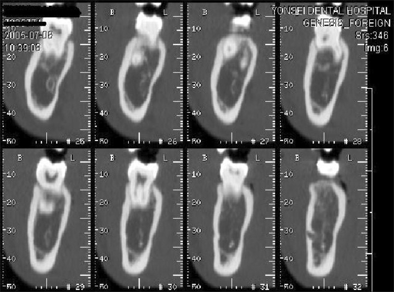
Figure 1 Representative computed tomography (CT) image of the transaxial slices of the mandibular body, as displayed by the PiView STAR program (INFINITT, Seoul, Korea).
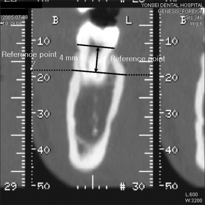
Figure 2 Reference points placed on an image of the reference site.
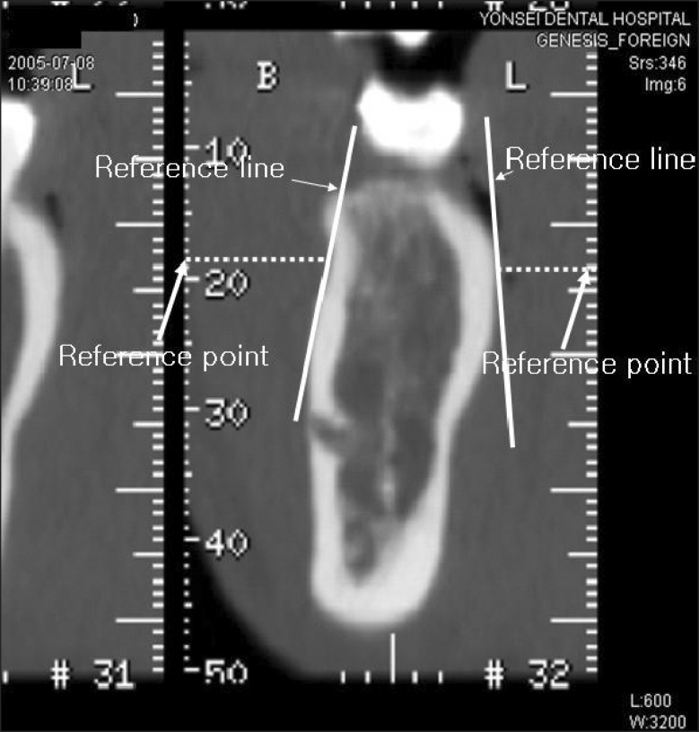
Figure 3 Reference lines placed on the measurement site.
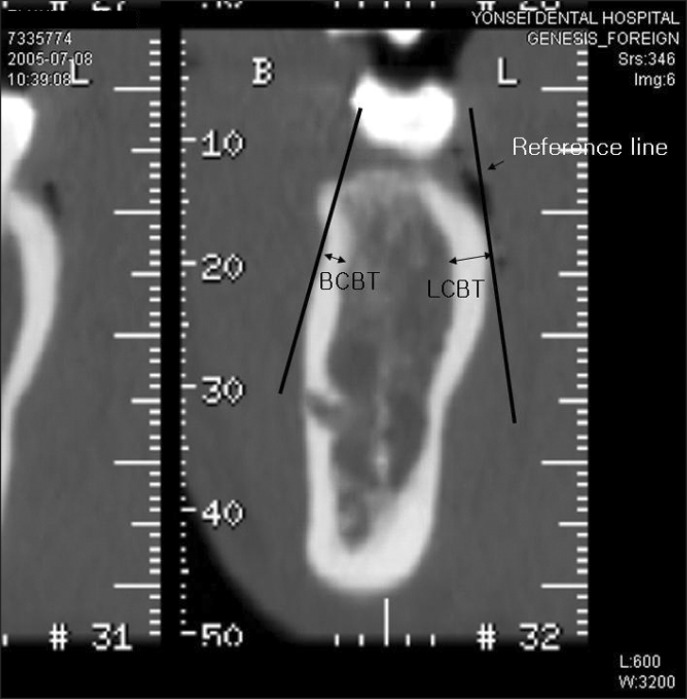
Figure 4 Measurement of buccal cortical bone thickness (BCBT) and lingual cortical bone thickness (LCBT).
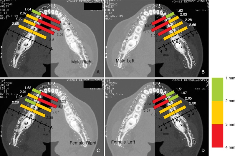
Figure 5 Average cortical bone thickness for (A) male right; (B) male left; (C) female right; (D) female left.
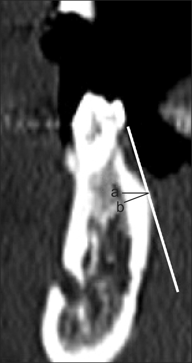
Figure 6 Cortical bone thickness from (a) the axial plane and (b) the transaxial plane as reference planes.
Cited by 3 articles
-
Three-dimensional evaluation of maxillary anterior alveolar bone for optimal placement of miniscrew implants
Jin Hwan Choi, Hyung Seog Yu, Kee Joon Lee, Young Chel Park
Korean J Orthod. 2014;44(2):54-61. doi: 10.4041/kjod.2014.44.2.54.Miniscrews versus surgical archwires for intermaxillary fixation in adults after orthognathic surgery
Sieun Son, Seong Sik Kim, Woo-Sung Son, Yong-Il Kim, Yong-Deok Kim, Sang-Hun Shin
Korean J Orthod. 2015;45(1):3-12. doi: 10.4041/kjod.2015.45.1.3.Stress distributions in peri-miniscrew areas from cylindrical and tapered miniscrews inserted at different angles
Sung-Hwan Choi, Seong-Jin Kim, Kee-Joon Lee, Sang-Jin Sung, Youn-Sic Chun, Chung-Ju Hwang
Korean J Orthod. 2016;46(4):189-198. doi: 10.4041/kjod.2016.46.4.189.
Reference
-
1. Poggio PM, Incorvati C, Velo S, Carano A. "Safe zones": a guide for miniscrew positioning in the maxillary and mandibular arch. Angle Orthod. 2006; 76:191–197. PMID: 16539541.2. Hu KS, Kang MK, Kim TW, Kim KH, Kim HJ. Relationships between dental roots and surrounding tissues for orthodontic miniscrew installation. Angle Orthod. 2009; 79:37–45. PMID: 19123704.
Article3. Park HS, Jeong SH, Kwon OW. Factors affecting the clinical success of screw implants used as orthodontic anchorage. Am J Orthod Dentofacial Orthop. 2006; 130:18–25. PMID: 16849067.
Article4. Santiago RC, de Paula FO, Fraga MR, Picorelli Assis NM, Vitral RW. Correlation between miniscrew stability and bone mineral density in orthodontic patients. Am J Orthod Dentofacial Orthop. 2009; 136:243–250. PMID: 19651355.
Article5. Miyamoto I, Tsuboi Y, Wada E, Suwa H, Iizuka T. Influence of cortical bone thickness and implant length on implant stability at the time of surgery--clinical, prospective, biomechanical, and imaging study. Bone. 2005; 37:776–780. PMID: 16154396.
Article6. Wilmes B, Rademacher C, Olthoff G, Drescher D. Parameters affecting primary stability of orthodontic mini-implants. J Orofac Orthop. 2006; 67:162–174. PMID: 16736117.
Article7. Kim DH. Anatomical characteristics of the midpalatal suture area for miniscrew implantation using CT image [master's thesis]. 2003. Seoul: Yonsei University.8. Lee JS, Kim DH, Park YC, Kyung SH, Kim TK. The efficient use of midpalatal miniscrew implants. Angle Orthod. 2004; 74:711–714. PMID: 15529509.9. Kim HJ, Yun HS, Park HD, Kim DH, Park YC. Soft-tissue and cortical-bone thickness at orthodontic implant sites. Am J Orthod Dentofacial Orthop. 2006; 130:177–182. PMID: 16905061.
Article10. Waitzman AA, Posnick JC, Armstrong DC, Pron GE. Craniofacial skeletal measurements based on computed tomography: Part I. Accuracy and reproducibility. Cleft Palate Craniofac J. 1992; 29:112–117. PMID: 1571344.
Article11. Hanazawa T, Sano T, Seki K, Okano T. Radiologic measurements of the mandible: a comparison between CT-reformatted and conventional tomographic images. Clin Oral Implants Res. 2004; 15:226–232. PMID: 15008935.12. Spoor CF, Zonneveld FW, Macho GA. Linear measurements of cortical bone and dental enamel by computed tomography: applications and problems. Am J Phys Anthropol. 1993; 91:469–484. PMID: 8372936.
Article13. Masumoto T, Hayashi I, Kawamura A, Tanaka K, Kasai K. Relationships among facial type, buccolingual molar inclination, and cortical bone thickness of the mandible. Eur J Orthod. 2001; 23:15–23. PMID: 11296507.
Article14. Kim JH, Joo JY, Park YW, Cha BK, Kim SM. Study of maxillary cortical bone thickness for skeletal anchorage system in Korean. J Korean Assoc Oral Maxillofac Surg. 2002; 28:249–255.15. Park HS. An anatomical study using CT images for the implantation of micro-implants. Korean J Orthod. 2002; 32:435–441.16. Kim JS. Clinical study on the width of attached gingiva in the subjects with healthy gingiva, or early stage of gingivitis [master's thesis]. 1997. Seoul: Yonsei University.17. Voigt JP, Goran ML, Flesher RM. The width of lingual mandibular attached gingiva. J Periodontol. 1978; 49:77–80. PMID: 276595.
Article18. Linde J, Lang NP, Karring T. Clinical periodontology and implant dentistry. 2008. 5th ed. Oxford: Blackwell Munksgaard;p. 6–8.19. Kim HJ. The morphology of the mandibular canal and the structure of the compact and sponge bone in Korean adult mandibles [master's thesis]. 1993. Seoul: Yonsei University.20. Silvestrini Biavati A, Tecco S, Migliorati M, Festa F, Marzo G, Gherlone E, et al. Three-dimensional tomographic mapping related to primary stability and structural miniscrew characteristics. Orthod Craniofac Res. 2011; 14:88–99. PMID: 21457458.
Article21. Tsunori M, Mashita M, Kasai K. Relationship between facial types and tooth and bone characteristics of the mandible obtained by CT scanning. Angle Orthod. 1998; 68:557–562. PMID: 9851354.22. Ichim I, Kieser JA, Swain MV. Functional significance of strain distribution in the human mandible under masticatory load: numerical predictions. Arch Oral Biol. 2007; 52:465–473. PMID: 17137552.
Article23. Hirabayashi M, Motoyoshi M, Ishimaru T, Kasai K, Namura S. Stresses in mandibular cortical bone during mastication: biomechanical considerations using a three-dimensional finite element method. J Oral Sci. 2002; 44:1–6. PMID: 12058864.
Article
- Full Text Links
-
- Actions
-
Cited
- CITED
-
- Close
- Share
-
- Similar articles
-
- Temporary replacement of congenital missing incisors on mandible using temporary anchorage devices in growing patient: 2-year follow-up
- Effectiveness of anchorage with temporary anchorage devices during anterior maxillary tooth retraction: A randomized clinical trial
- Histological analysis on tissues around orthodontically intruded maxillary molars using temporary anchorage devices: A case report
- Cortical bone thickness and root proximity at mandibular interradicular sites: implications for orthodontic mini-implant placement
- Quantitative evaluation of cortical bone and soft tissue thickness in the mandible
