Temporary replacement of congenital missing incisors on mandible using temporary anchorage devices in growing patient: 2-year follow-up
- Affiliations
-
- 1Dental Clinic Center, Pusan National University Hosptial, Busan, Republic of Korea
- 2Department of Orthodontics, School of Dentistry, Pusan National University, Yangsan, Republic of Korea
- KMID: 2512122
- DOI: http://doi.org/10.14368/jdras.2020.36.4.272
Abstract
- Agenesis of permanent tooth in adolescent patients can be treated either by orthodontic treatment for space closure or by main-taining the space until implant restoration can be carried out in adult. However, gradual atrophy of alveolar bone width makes it dif-ficult to restore the prosthesis in the future or may cause unaesthetic results. Therefore, maintaining of not only the missing space but also the alveolar bone width should be considered. This case is a treatment whereby a temporary replacement of missing 2 mandibular incisors in adolescent patient was carried out using 2 temporary anchorage devices (TADs). Two TADs were placed hori-zontally 2 - 3 mm below the top of alveolar ridge, and fixed with artificial teeth by stainless steel wires extended. During the 2 year follow-up, neither gingival inflammation nor loss of the TADs have occurred. In the radiographic evaluation, the growth of the adja-cent alveolar bone was not inhibited, and the width of the alveolar bone was maintained.
Figure
-
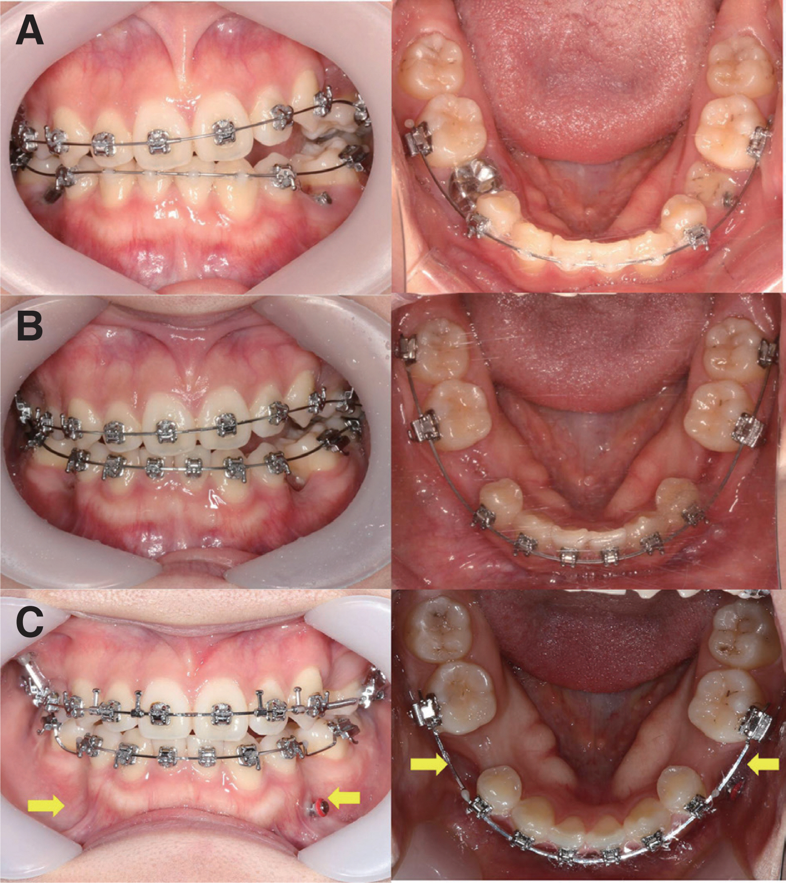
Fig. 1 Clinical pictures (A - C) of a patient suffering from agenesis of both mandibular second premolars and buccal open bite on left side. (A) Both mandibular second primary molars were ankylosed and infraocclusion. (B) Extraction of ankylosed primary molars. And 1 month later, temporary anchorage devices (TADs) was inserted on left side for using as an anchorage for inter-maxillary elastics. (C) After 1 year, the width and height of the alveolar bone were better retained with TADs than without TADs (yellow arrows).

Fig. 2 Pre-treatment intraoral (A) and extraoral (B) photographs. (A) 2 Mandibular anterior teeth were congenitally missing. (B) Prominent chin and concave profile.
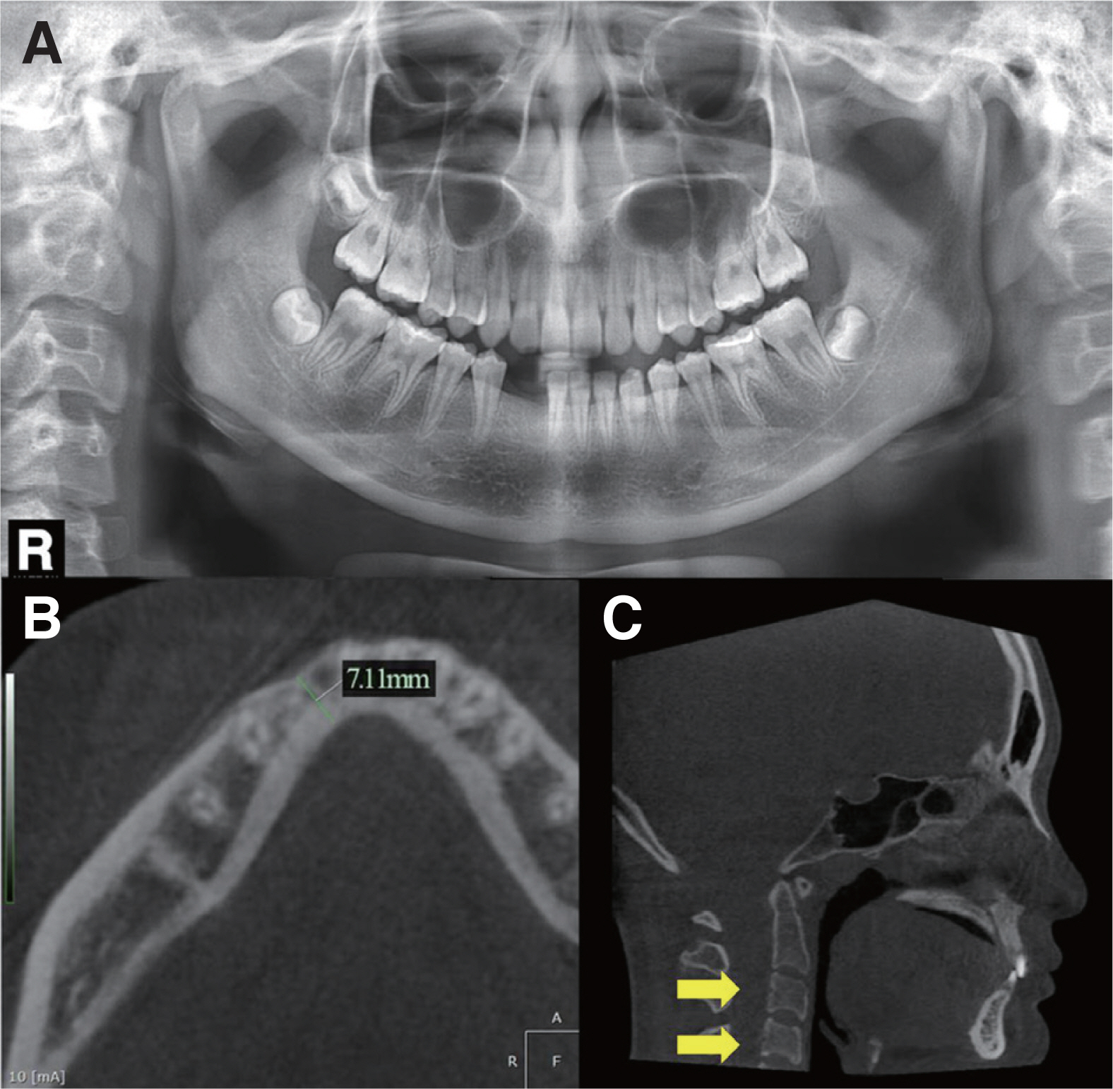
Fig. 3 Pre-treatment panoramic (A) and CBCT (B, C) radiographs. (A) Panoramic radiograph. (B) Cone-beam computed tomograph (axial view) : Alveolar bone width (most narrow area) was 7.11 mm. (C) Inferior borders of the second, third and fourth vertebra’s bodys were concave and body’s shapes were rectangular (yellow arrows). Therefore, she was estimated on CVM stage 4 (after growth peak period).

Fig. 4 Temporary anchorage devices (TADs) were placed at 2 - 3 mm under the alveolar crest and temporary artificial teeth with loop were placed.

Fig. 5 Intraoral photographs of 2-year follow-up. No inflammation of the soft tissues around the TADs was detected.
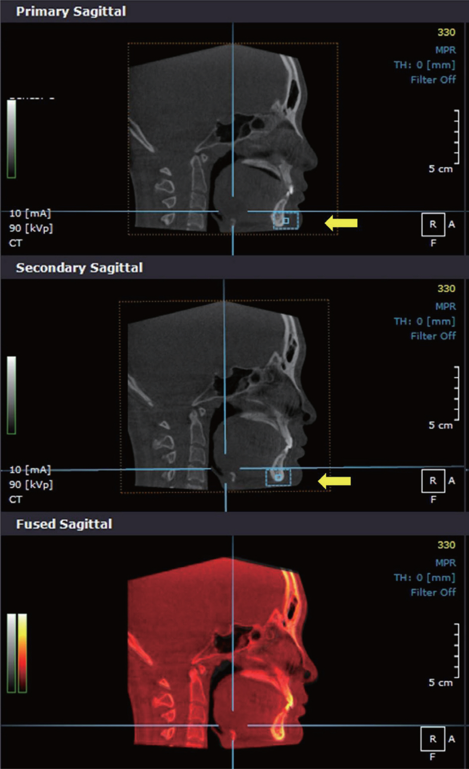
Fig. 6 Superimpositions of pre-treatment and post-treatment CBCTs. Chin and symphysis of mandible excluding teeth (yellow arrows, dotted rectangles) were used as a stabilizing structure for superimposition.
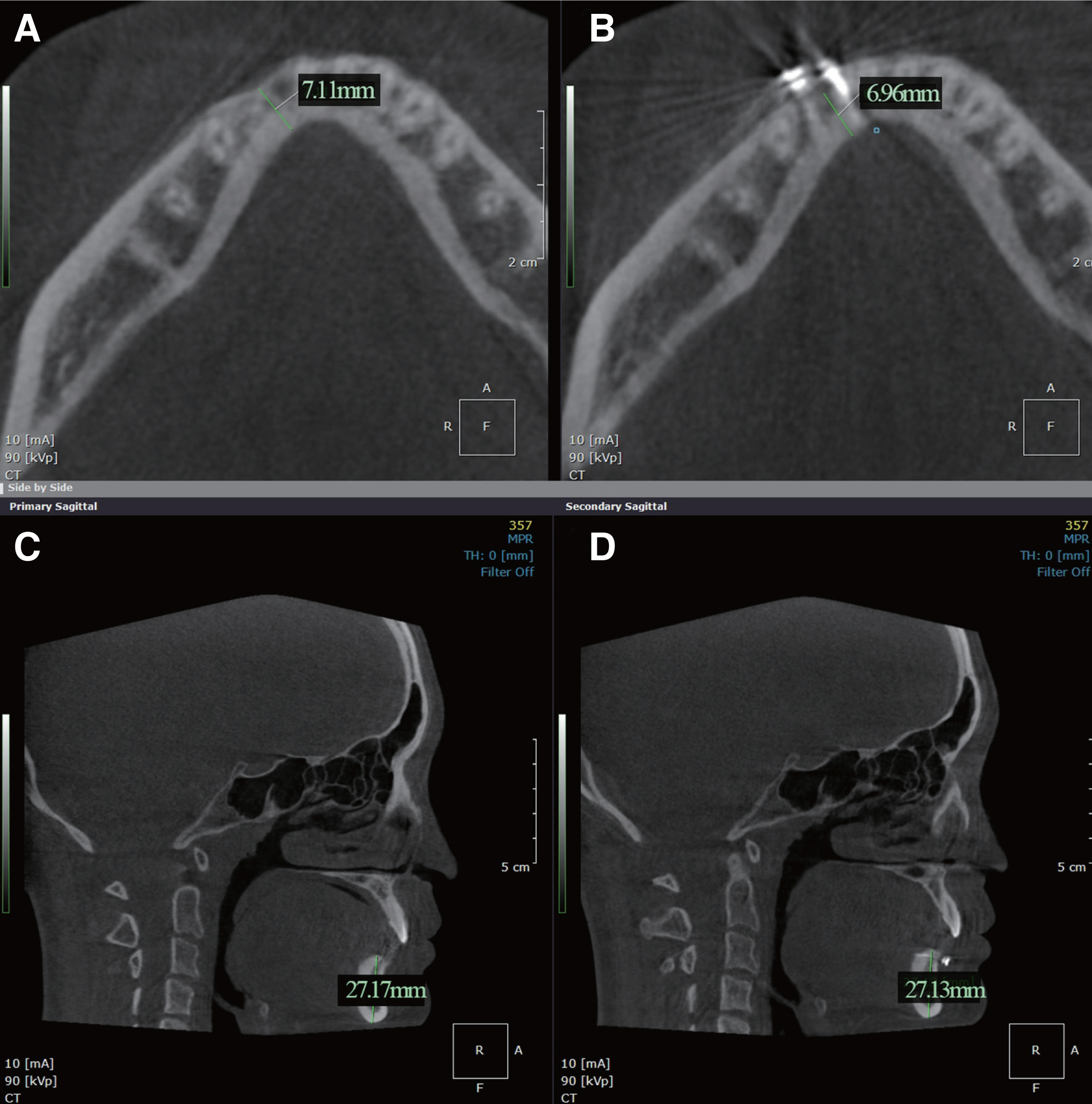
Fig. 7 Comparisons of pre-treatment (A, C) and post-treatment (B, D). Alveolar bone width (most narrow area, CBCT axial view): (A) 7.11 mm, (B) 6.96 mm. Alveolar bone height (CBCT sagittal view): (C) 27.17 mm, (D) 27.13 mm. No significant changes were observed.
Reference
-
References
1. Goya HA, Tanaka S, Maeda T, Akimoto Y. 2008; An orthopantomographic study of hypodontia in permanent teeth of Japanese pediatric patients. J Oral Sci. 50:143–50. DOI: 10.2334/josnusd.50.143. PMID: 18587203.2. Polder BJ, Van't Hof MA, Van der Linden FP, Kuijpers-Jagtman AM. 2004; A meta-analysis of the prevalence of dental agenesis of permanent teeth. Community Dent Oral Epidemiol. 32:217–26. DOI: 10.1111/j.1600-0528.2004.00158.x. PMID: 15151692.3. Korean Academy of Pediatric Dentistry. 2014. Pediatric adolescent dentistry. 5th ed. Yenang Inc;Seoul: p. 566–9.4. Kokich VG, Kokich VO. 2006; Congenitally missing mandibular second premolars: clinical options. Am J Orthod Dentofacial Orthop. 130:437–44. DOI: 10.1016/j.ajodo.2006.05.025. PMID: 17045142.5. Thilander B, Odman J, Lekholm U. 2001; Orthodontic aspects of the use of oral implants in adolescents: a 10-year follow-up study. Eur J Orthod. 23:715–31. DOI: 10.1093/ejo/23.6.715. PMID: 11890067.6. Fudalej P, Kokich VG, Leroux B. 2007; Determining the cessation of vertical growth of the craniofacial structures to facilitate placement of singletooth implants. Am J Orthod Dentofacial Orthop. 131 Suppl 4:S59–67. DOI: 10.1016/j.ajodo.2006.07.022. PMID: 17448387.7. Olsen TM, Kokich VG Sr. 2010; Postorthodontic root approximation after opening space for maxillary lateral incisor implants. Am J Orthod Dentofacial Orthop. 137:158.e1–8. DOI: 10.1016/j.ajodo.2009.08.024. PMID: 20152659.8. Jeong DM, Choi B, Choo H, Kim JH, Chung KR, Kim SH. 2011; Novel application of the 2-piece orthodontic C-implant for temporary crown restoration after orthodontic treatment. Am J Orthod Dentofacial Orthop. 140:569–79. DOI: 10.1016/j.ajodo.2009.10.051. PMID: 21967946.9. Kokich VG, Swift EJ Jr. 2011; Temporary restoration of maxillary lateral incisor implant sites. J Esthet Restor Dent. 23:136–7. DOI: 10.1111/j.1708-8240.2011.00440.x. PMID: 21649825.10. Bollero P, Fazio V, Pavoni C, Cordaro M, Cozza P, Lione R. 2018; Titanium alloy vs. stainless steel miniscrews: an in vivo split-mouth study. Eur Rev Med Pharmacol Sci. 22:2191–8. DOI: 10.26355/eurrev_201804_14803. PMID: 29762818.11. Melsen B, Huja SS, Chien HH, Dalstra M. 2015; Alveolar bone preservation subsequent to miniscrew implant placement in a canine model. Orthod Craniofac Res. 18:77–85. DOI: 10.1111/ocr.12058. PMID: 25403977.12. Ciarlantini R, Melsen B. 2017; Semipermanent replacement of missing maxillary lateral incisors by miniimplant retained pontics: A follow-up study. Am J Orthod Dentofacial Orthop. 151:989–94. DOI: 10.1016/j.ajodo.2016.12.012. PMID: 28457277.13. Hunter CJ. 1966; The correlation of facial growth with body height and skeletal maturation at adolescence. Angle Orthod. 36:44–54. DOI: 10.1043/0003-3219(1966)036<0044:TCOFGW>2.0.CO;2. PMID: 5218761.14. Baccetti T, Franchi L, McNamara JA Jr. 2005; The cervical vertebral maturation (CVM) method for the assessment of optimal treatment timing in dentofacial orthopedics. Semin Orthod. 11:119–29. DOI: 10.1053/j.sodo.2005.04.005.15. Nguyen T, Cevidanes L, Franchi L, Ruellas A, Jackson T. 2018; Three-dimensional mandibular regional superimposition in growing patients. Am J Orthod Dentofac Orthop. 153:747–54. DOI: 10.1016/j.ajodo.2017.07.026. PMID: 29706223.16. Johnson K. 1969; A study of the dimensional changes occurring in the maxilla following tooth extraction. Aust Dent J. 14:241–4. DOI: 10.1111/j.1834-7819.1969.tb06001.x. PMID: 5259350.17. Schropp L, Wenzel A, Kostopoulos L, Karring T. 2003; Bone healing and soft tissue contour changes following single-tooth extraction: a clinical and radiographic 12-month prospective study. Int J Periodontics Restorative Dent. 23:313–23. PMID: 12956475.18. Ostler MS, Kokich VG. 1994; Alveolar ridge changes in patients congenitally missing mandibular second premolars. J Prosthet Dent. 71:144–9. DOI: 10.1016/0022-3913(94)90022-1. PMID: 8126668.19. Bertl K, Bertl MH, Heimel P, Burt M, Gahleitner A, Stavropoulos A, Ulm C. 2018; Alveolar bone resorption after primary tooth loss has a negative impact on straightforward implant installation in patients with agenesis of the lower second premolar. Clin Oral Implants Res. 29:155–63. DOI: 10.1111/clr.13033. PMID: 28736870.20. Williams P, Travess H, Sandy J. 2004; The use of osseointegrated implants in orthodontic patiens: I. Implants and their use in children. Dent Update. 31:287–90. DOI: 10.12968/denu.2004.31.5.287. PMID: 15242262.21. Gill DS, Jones S, Hobkirk J, Bassi S, Hemmings K, Goodman J. 2008; Counselling patients with hypodontia. Dent Update. 35:344–52. DOI: 10.12968/denu.2008.35.5.344. PMID: 18605529.
- Full Text Links
-
- Actions
-
Cited
- CITED
-
- Close
- Share
-
- Similar articles
-
- Improvements of facial profile and smile aesthetic using temporary anchorage devices and botulinum toxin: a case report
- Effectiveness of anchorage with temporary anchorage devices during anterior maxillary tooth retraction: A randomized clinical trial
- Nonextraction treatment of Class II division 2 in an adult patient using microimplant anchorage (MIA)
- Evaluation of mandibular cortical bone thickness for placement of temporary anchorage devices (TADs)
- Study Of Maxillary Cortical Bone Thickness For Skeletal Anchorage System In Korean
