Primary malignant rhabdoid tumor of greater omentum in 10-year-old girl
- Affiliations
-
- 1Department of Pediatric Surgery, Inje University Haeundae Paik Hospital, Busan, Korea. namsh@paik.ac.kr
- 2Department of Pediatrics, Inje University Haeundae Paik Hospital, Busan, Korea.
- 3Department of Pathology, Inje University Haeundae Paik Hospital, Busan, Korea.
- KMID: 2266907
- DOI: http://doi.org/10.4174/astr.2014.86.1.50
Abstract
- Contrary to metastatic tumors of the omentum, primary tumors of the omentum are very rare. A 10-year-old girl presented with low abdominal pain. Imaging studies showed a multiseptated hemorrhagic tumor. The mass from the omentum was removed completely and confirmed as a malignant rhabdoid tumor. Despite aggressive chemotherapy, she died after 9 months due to disease progression. We report one case of primary malignant rhabdoid tumor of the omentum for the first time.
Keyword
Figure
-

Fig. 1 The computed tomography scan showed lobulating contoured hemorrhagic necrotic tumor (9 cm × 6 cm × 6.5 cm) with peripheral irregular enhancement in lower abdomen and pelvis and some amount of hemoperitoneum in cul-de-sac.
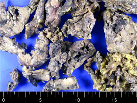
Fig. 2 On formalin-fixed specimen, multinodular solid masses showed tan gray and fleshy cut surface. Multifocal hemorrhage and necrosis were seen.
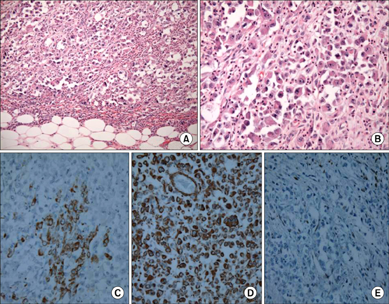
Fig. 3 Microscopic findings: tumor cells showed characteristic findings of "rhabdoid" cells such as infiltrative border (A: H&E, ×200), eccentric nuclei, prominent nucleoli, and characteristic eosinophilic inclusion or globules in abundant cytoplasm (B: H&E, ×400) in nested tumor cells. Immunohistochemically neoplastic cells expressed cytokeratin (C, ×400), and vimentin (D, ×400). Importantly neoplastic cells revealed absence of nuclear expression of INI 1 (E, ×200).
Reference
-
1. Ishida H, Ishida J. Primary tumours of the greater omentum. Eur Radiol. 1998; 8:1598–1601.2. Haaga JR. The peritoneum and mesentery. In : Haaga JR, Lanzieri CF, editors. Computed tomography and magnetic resonance imaging of the whole body. St. Louis: Mosby;1994. p. 1244–1291.3. Sompayrac SW, Mindelzun RE, Silverman PM, Sze R. The greater omentum. AJR Am J Roentgenol. 1997; 168:683–687.4. Sigauke E, Rakheja D, Maddox DL, Hladik CL, White CL, Timmons CF, et al. Absence of expression of SMARCB1/INI1 in malignant rhabdoid tumors of the central nervous system, kidneys and soft tissue: an immunohistochemical study with implications for diagnosis. Mod Pathol. 2006; 19:717–725.5. Evans KJ, Miller Q, Kline AL. Solid omental tumors [Internet]. New York: WebMD LLC;c1994-2013. cited 2011 Nov 28. Available from: http://emedicine.medscape.com/article/193622.6. Reinhard H, Reinert J, Beier R, Furtwangler R, Alkasser M, Rutkowski S, et al. Rhabdoid tumors in children: prognostic factors in 70 patients diagnosed in Germany. Oncol Rep. 2008; 19:819–823.7. Leong FJ, Leong AS. Malignant rhabdoid tumor in adults: heterogenous tumors with a unique morphological phenotype. Pathol Res Pract. 1996; 192:796–807.8. Amrikachi M, Ro JY, Ordonez NG, Ayala AG. Adenocarcinomas of the gastrointestinal tract with prominent rhabdoid features. Ann Diagn Pathol. 2002; 6:357–363.9. Chung CJ, Cammoun D, Munden M. Rhabdoid tumor of the kidney presenting as an abdominal mass in a newborn. Pediatr Radiol. 1990; 20:562–563.10. Hilden JM, Meerbaum S, Burger P, Finlay J, Janss A, Scheithauer BW, et al. Central nervous system atypical teratoid/rhabdoid tumor: results of therapy in children enrolled in a registry. J Clin Oncol. 2004; 22:2877–2884.
