Characterization of nestin expression in astrocytes in the rat hippocampal CA1 region following transient forebrain ischemia
- Affiliations
-
- 1Department of Anatomy and Cell Death Disease Research Center, The Catholic University of Korea College of Medicine, Seoul, Korea. munylee@catholic.ac.kr
- KMID: 2263105
- DOI: http://doi.org/10.5115/acb.2013.46.2.131
Abstract
- Recent studies have suggested that nestin facilitates cellular structural remodeling in vasculature-associated cells in response to ischemic injury. The current study was designed to investigate the potential role of post-ischemic nestin expression in parenchymal astrocytes. With this aim, we characterized ischemia-induced nestin expression in the CA1 hippocampal region, an area that undergoes a delayed neuronal death, followed by a lack of neuronal generation after transient forebrain ischemia. Virtually all of the nestin-positive cells in the ischemic CA1 hippocampus were reactive astrocytes. However, induction of nestin expression did not correlate simply with astrogliosis, but rather showed characteristic time- and strata-dependent expression patterns. Nestin induction in astrocytes of the pyramidal cell layer was rapid and transient, while a long-lasting induction of nestin was observed in astrocytes located in the CA1 dendritic subfields, such as the stratum oriens and radiatum, until at least day 28 after ischemia. There was no detectable expression in the stratum lacunosum moleculare despite the evident astroglial reaction. Almost all of the nestin-positive cells also expressed a transcription factor for neural/glial progenitors, i.e., Sox-2 or Sox-9, and some cells were also positive for Ki-67. However, all of the nestin-positive astrocytes expressed the calcium-binding protein S100beta, which is known to be expressed in a distinct, post-mitotic astrocyte population. Thus, our data indicate that in the ischemic CA1 hippocampus, nestin expression was induced in astroglia that were becoming reactive, but not in a progenitor/stem cell population, suggesting that nestin may allow for the structural remodeling of these cells in response to ischemic injury.
MeSH Terms
Figure
-
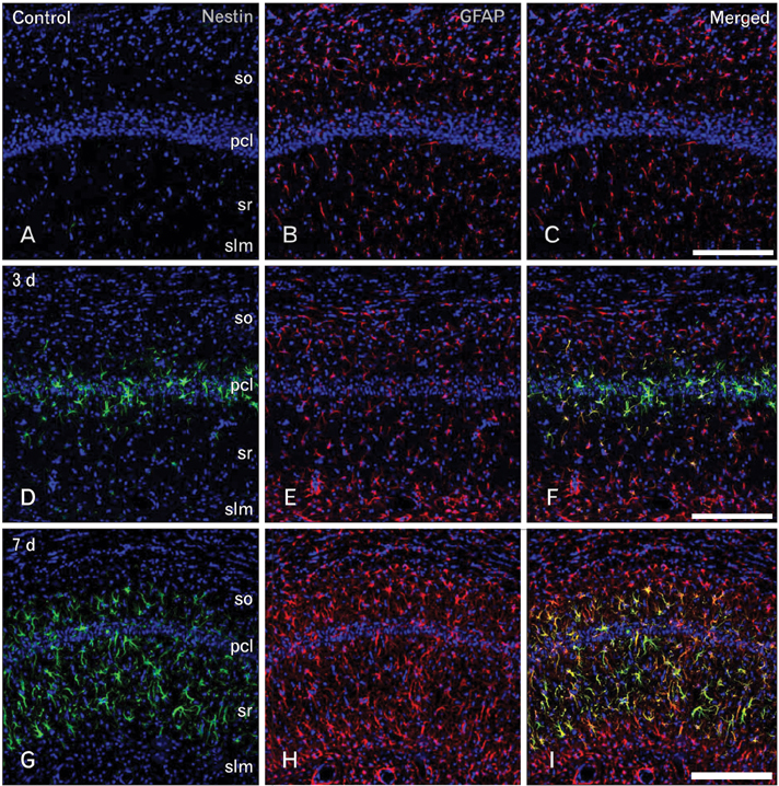
Fig. 1 Spatiotemporal relationship between nestin and glial fibrillary acidic protein (GFAP) in the hippocampal CA1 region in the early phase following transient forebrain ischemia. (A-C) No specific staining for nestin was detected in the hippocampal CA1 region of sham-operated rats, while GFAP-positive astrocytes showed thin glial processes. pcl, pyramidal cell layer; slm, stratum lacunosum moleculare; so, stratum oriens; sr, stratum radiatum. (D-F) At day 3 after ischemia, significant induction of nestin expression was detected in the CA1 pyramidal cell layer, while a rather uniform upregulation of GFAP immunoreactivity was observed in all laminae of the CA1 region. (G-I) At 7 days, GFAP immunoreactivity was markedly increased in all strata of the CA1 region, while nestin expression was more pronounced in the CA1 dendritic layers and absent in the stratum lacunosum moleculare. Scale bars in (C, F, I)=200 µm (A-I).

Fig. 2 Spatiotemporal relationship between nestin and glial fibrillary acidic protein (GFAP) in the hippocampal CA1 region during the later phase following transient forebrain ischemia. (A-C) At 14 days after ischemia, GFAP-positive astrocytes were distributed in all strata of the hippocampal CA1 region, while nestin expression was localized in the CA1 dendritic layers including the strata radiatum (sr) and oriens (so), but was rare in the pyramidal cell layer (pcl) and in the stratum lacunosum moleculare (slm). Over the chronic interval of 21 days (D-F) and 28 days (G-I), the labeling intensity and patterns of GFAP staining were similar to those at day 14. Nestin expression was still observed in the CA1 dendritic layers at 21 days after ischemia, while it remained only in the stratum radiatum at 28 days. Note the absence of nestin expression in the stratum lacunosum moleculare. Scale bars in (C, F, I)=200 µm (A-I).
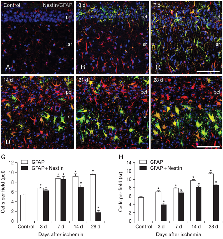
Fig. 3 Spatiotemporal distribution of nestin-positive astrocytes in the CA1 hippocampus of control (A) and ischemic rats (B-F). Nestin expression appeared in the pyramidal cell layer (pcl) by 3 days (B), and extended into the stratum radiatum (sr) by 7 days (C). By 14 days after ischemia (D), nestin expression was more pronounced in the stratum radiatum, and further increased in this stratum at 21 days (E) and 28 days (F). Note that nestin-positive astrocytes were rarely observed in the pyramidal cell layer by 14 days, despite the evident astroglial reaction. (G, H) Temporal profile of glial fibrillary acidic protein (GFAP) single-labeled cells and nestin/GFAP double-labeled cells in the pyramidal cell layer (G) and stratum radiatum (H) of the CA1 region after transient forebrain ischemia. The data are expressed as the mean±SD; *P<0.01 from the Bonferroni test with each time point compared to sham-operated controls. Scale bars in (C, F)=100 µm (A-F).

Fig. 4 Characterization of nestin-positive cells in the CA1 hippocampal region after transient forebrain ischemia. (A-F) Double-labeling with nestin and Ki-67 showed that some nestin-positive cells exhibited Ki-67-labeled nuclei (arrowheads) on days 3 (A-C) and 14 (D-F) after ischemia. pcl, pyramidal cell layer; sr, stratum radiatum. (G-L) Double-labeling with nestin and the transcription factors Sox-2 (G-I) or Sox-9 (J-L). Note that almost all nestin-positive cells expressed Sox-2- or Sox-9-labeled nuclei at days 3 (H, K) and 14 (I, L) after ischemia. Also note that Sox-2- or Sox-9-positive nuclei were also observed in the control hippocampus (G, J), where no nestin expression was observed. Scale bars in (C, F, I, L)=100 µm (A-L).

Fig. 5 Relationship between S100β and nestin (A, B, D, E) or glial fibrillary acidic protein (GFAP) (C, F) in the CA1 hippocampal region after transient forebrain ischemia. All of the nestin-positive cells expressed S100β, corresponding to only a small fraction of all of the S100β-positive cells at all time points examined. Note that virtually all of the S100β-positive cells in the ischemic CA1 region were GFAP-positive astrocytes. Scale bars in (C, F)=100 µm (A-F).
Cited by 1 articles
-
Role of agmatine in the application of neural progenitor cell in central nervous system diseases: therapeutic potentials and effects
Renée Kosonen, Sumit Barua, Jong Youl Kim, Jong Eun Lee
Anat Cell Biol. 2021;54(2):143-151. doi: 10.5115/acb.21.089.
Reference
-
1. Hockfield S, McKay RD. Identification of major cell classes in the developing mammalian nervous system. J Neurosci. 1985. 5:3310–3328.2. Lendahl U, Zimmerman LB, McKay RD. CNS stem cells express a new class of intermediate filament protein. Cell. 1990. 60:585–595.3. Morshead CM, Reynolds BA, Craig CG, McBurney MW, Staines WA, Morassutti D, Weiss S, van der Kooy D. Neural stem cells in the adult mammalian forebrain: a relatively quiescent subpopulation of subependymal cells. Neuron. 1994. 13:1071–1082.4. Sejersen T, Lendahl U. Transient expression of the intermediate filament nestin during skeletal muscle development. J Cell Sci. 1993. 106(Pt 4):1291–1300.5. Kachinsky AM, Dominov JA, Miller JB. Intermediate filaments in cardiac myogenesis: nestin in the developing mouse heart. J Histochem Cytochem. 1995. 43:843–847.6. Toma JG, Akhavan M, Fernandes KJ, Barnabé-Heider F, Sadikot A, Kaplan DR, Miller FD. Isolation of multipotent adult stem cells from the dermis of mammalian skin. Nat Cell Biol. 2001. 3:778–784.7. Zulewski H, Abraham EJ, Gerlach MJ, Daniel PB, Moritz W, Müller B, Vallejo M, Thomas MK, Habener JF. Multipotential nestin-positive stem cells isolated from adult pancreatic islets differentiate ex vivo into pancreatic endocrine, exocrine, and hepatic phenotypes. Diabetes. 2001. 50:521–533.8. Kim SY, Lee SH, Kim BM, Kim EH, Min BH, Bendayan M, Park IS. Activation of nestin-positive duct stem (NPDS) cells in pancreas upon neogenic motivation and possible cytodifferentiation into insulin-secreting cells from NPDS cells. Dev Dyn. 2004. 230:1–11.9. Barres BA. A new role for glia: generation of neurons. Cell. 1999. 97:667–670.10. Alvarez-Buylla A, García-Verdugo JM, Tramontin AD. A unified hypothesis on the lineage of neural stem cells. Nat Rev Neurosci. 2001. 2:287–293.11. Tamamaki N, Nakamura K, Okamoto K, Kaneko T. Radial glia is a progenitor of neocortical neurons in the developing cerebral cortex. Neurosci Res. 2001. 41:51–60.12. Campbell K, Götz M. Radial glia: multi-purpose cells for vertebrate brain development. Trends Neurosci. 2002. 25:235–238.13. Mori T, Buffo A, Götz M. The novel roles of glial cells revisited: the contribution of radial glia and astrocytes to neurogenesis. Curr Top Dev Biol. 2005. 69:67–99.14. Gilyarov AV. Nestin in central nervous system cells. Neurosci Behav Physiol. 2008. 38:165–169.15. Ehninger D, Kempermann G. Neurogenesis in the adult hippocampus. Cell Tissue Res. 2008. 331:243–250.16. von Bohlen und Halbach O. Immunohistological markers for proliferative events, gliogenesis, and neurogenesis within the adult hippocampus. Cell Tissue Res. 2011. 345:1–19.17. Rosen GD, Sherman GF, Galaburda AM. Radial glia in the neocortex of adult rats: effects of neonatal brain injury. Brain Res Dev Brain Res. 1994. 82:127–135.18. Lin RC, Matesic DF, Marvin M, McKay RD, Brüstle O. Re-expression of the intermediate filament nestin in reactive astrocytes. Neurobiol Dis. 1995. 2:79–85.19. Duggal N, Schmidt-Kastner R, Hakim AM. Nestin expression in reactive astrocytes following focal cerebral ischemia in rats. Brain Res. 1997. 768:1–9.20. Li Y, Chopp M. Temporal profile of nestin expression after focal cerebral ischemia in adult rat. Brain Res. 1999. 838:1–10.21. Krum JM, Rosenstein JM. Transient coexpression of nestin, GFAP, and vascular endothelial growth factor in mature reactive astroglia following neural grafting or brain wounds. Exp Neurol. 1999. 160:348–360.22. Pforte C, Henrich-Noack P, Baldauf K, Reymann KG. Increase in proliferation and gliogenesis but decrease of early neurogenesis in the rat forebrain shortly after transient global ischemia. Neuroscience. 2005. 136:1133–1146.23. Lim DA, Huang YC, Alvarez-Buylla A. The adult neural stem cell niche: lessons for future neural cell replacement strategies. Neurosurg Clin N Am. 2007. 18:81–92.24. Ihrie RA, Alvarez-Buylla A. Cells in the astroglial lineage are neural stem cells. Cell Tissue Res. 2008. 331:179–191.25. Kirino T. Delayed neuronal death in the gerbil hippocampus following ischemia. Brain Res. 1982. 239:57–69.26. Pulsinelli WA, Brierley JB, Plum F. Temporal profile of neuronal damage in a model of transient forebrain ischemia. Ann Neurol. 1982. 11:491–498.27. Smith ML, Auer RN, Siesjö BK. The density and distribution of ischemic brain injury in the rat following 2-10 min of forebrain ischemia. Acta Neuropathol. 1984. 64:319–332.28. Tonchev AB, Yamashima T. Differential neurogenic potential of progenitor cells in dentate gyrus and CA1 sector of the postischemic adult monkey hippocampus. Exp Neurol. 2006. 198:101–113.29. Pulsinelli WA, Brierley JB. A new model of bilateral hemispheric ischemia in the unanesthetized rat. Stroke. 1979. 10:267–272.30. Lee MY, Shin SL, Choi YS, Kim EJ, Cha JH, Chun MH, Lee SB, Kim SY. Transient upregulation of osteopontin mRNA in hippocampus and striatum following global forebrain ischemia in rats. Neurosci Lett. 1999. 271:81–84.31. Paxinos G, Tork I, Tecott LH, Valentino KL. Altas of the developing rat brain. 2008. San Diego: Academic Press.32. Liu J, Bartels M, Lu A, Sharp FR. Microglia/macrophages proliferate in striatum and neocortex but not in hippocampus after brief global ischemia that produces ischemic tolerance in gerbil brain. J Cereb Blood Flow Metab. 2001. 21:361–373.33. Stolt CC, Lommes P, Sock E, Chaboissier MC, Schedl A, Wegner M. The Sox9 transcription factor determines glial fate choice in the developing spinal cord. Genes Dev. 2003. 17:1677–1689.34. Chu MS, Chang CF, Yang CC, Bau YC, Ho LL, Hung SC. Signalling pathway in the induction of neurite outgrowth in human mesenchymal stem cells. Cell Signal. 2006. 18:519–530.35. Domowicz MS, Henry JG, Wadlington N, Navarro A, Kraig RP, Schwartz NB. Astrocyte precursor response to embryonic brain injury. Brain Res. 2011. 1389:35–49.36. Seri B, García-Verdugo JM, McEwen BS, Alvarez-Buylla A. Astrocytes give rise to new neurons in the adult mammalian hippocampus. J Neurosci. 2001. 21:7153–7160.37. Filippov V, Kronenberg G, Pivneva T, Reuter K, Steiner B, Wang LP, Yamaguchi M, Kettenmann H, Kempermann G. Subpopulation of nestin-expressing progenitor cells in the adult murine hippocampus shows electrophysiological and morphological characteristics of astrocytes. Mol Cell Neurosci. 2003. 23:373–382.38. Tanaka H, Araki M, Masuzawa T. Reaction of astrocytes in the gerbil hippocampus following transient ischemia: immunohistochemical observations with antibodies against glial fibrillary acidic protein, glutamine synthetase, and S-100 protein. Exp Neurol. 1992. 116:264–274.39. Ordy JM, Wengenack TM, Bialobok P, Coleman PD, Rodier P, Baggs RB, Dunlap WP, Kates B. Selective vulnerability and early progression of hippocampal CA1 pyramidal cell degeneration and GFAP-positive astrocyte reactivity in the rat four-vessel occlusion model of transient global ischemia. Exp Neurol. 1993. 119:128–139.40. Schmidt-Kastner R, Szymas J. Immunohistochemistry of glial fibrillary acidic protein, vimentin and S-100 protein for study of astrocytes in hippocampus of rat. J Chem Neuroanat. 1990. 3:179–192.41. Witter MP, Amaral DG. Paxinos G, editor. Hippocampal formation. The Rat Nervous System. 2004. 3rd ed. San Diego: Elsevier Academic Press;635–704.42. Herrmann H, Aebi U. Intermediate filaments and their associates: multi-talented structural elements specifying cytoarchitecture and cytodynamics. Curr Opin Cell Biol. 2000. 12:79–90.43. Wang N, Stamenović D. Contribution of intermediate filaments to cell stiffness, stiffening, and growth. Am J Physiol Cell Physiol. 2000. 279:C188–C194.44. Chen LW, Zhang JP, Kwok-Yan Shum D, Chan YS. Localization of nerve growth factor, neurotrophin-3, and glial cell line-derived neurotrophic factor in nestin-expressing reactive astrocytes in the caudate-putamen of 1-methyl-4-phenyl-1,2,3,6- tetrahydropyridine-treated C57/Bl mice. J Comp Neurol. 2006. 497:898–909.45. Calderone A. Nestin+ cells and healing the infarcted heart. Am J Physiol Heart Circ Physiol. 2012. 302:H1–H9.46. Petito CK, Morgello S, Felix JC, Lesser ML. The two patterns of reactive astrocytosis in postischemic rat brain. J Cereb Blood Flow Metab. 1990. 10:850–859.47. Shin YJ, Kim HL, Park JM, Cho JM, Kim SY, Lee MY. Characterization of nestin expression and vessel association in the ischemic core following focal cerebral ischemia in rats. Cell Tissue Res. 2013. 351:383–395.48. Nakatomi H, Kuriu T, Okabe S, Yamamoto S, Hatano O, Kawahara N, Tamura A, Kirino T, Nakafuku M. Regeneration of hippocampal pyramidal neurons after ischemic brain injury by recruitment of endogenous neural progenitors. Cell. 2002. 110:429–441.49. Schmidt W, Reymann KG. Proliferating cells differentiate into neurons in the hippocampal CA1 region of gerbils after global cerebral ischemia. Neurosci Lett. 2002. 334:153–156.50. Salazar-Colocho P, Lanciego JL, Del Rio J, Frechilla D. Ischemia induces cell proliferation and neurogenesis in the gerbil hippocampus in response to neuronal death. Neurosci Res. 2008. 61:27–37.51. Buffo A, Rite I, Tripathi P, Lepier A, Colak D, Horn AP, Mori T, Gotz M. Origin and progeny of reactive gliosis: A source of multipotent cells in the injured brain. Proc Natl Acad Sci U S A. 2008. 105:3581–3586.
- Full Text Links
-
- Actions
-
Cited
- CITED
-
- Close
- Share
-
- Similar articles
-
- Increase of peroxynitrite production in the rat brain following transient forebrain ischemia
- Postischemic Treatment with Aminoguanidine Inhibits Peroxynitrite Production in the Rat Hippocampus Following Transient Forebrain Ischemia
- The Effects of Propofol and Sevoflurane on Neuronal Apoptosis and Bcl-2 Family Protein Expression after Transient Forebrain Ischemia in the Rat
- Temporal changes in mammalian target of rapamycin (mTOR) and phosphorylated-mTOR expressions in the hippocampal CA1 region of rat with vascular dementia
- Effect of sedative dose of propofol on neuronal damage after transient forebrain ischemia in Mongolian gerbils
