Relationship between serum interleukin-31/25-hydroxyvitamin D levels and the severity of atopic dermatitis in children
- Affiliations
-
- 1Department of Pediatrics, Kangbuk Samsung Hospital, Sungkyunkwan University School of Medicine, Seoul, Korea. jy7.shim@samsung.com
- KMID: 2218627
- DOI: http://doi.org/10.4168/aard.2015.3.6.396
Abstract
- PURPOSE
Atopic dermatitis (AD) is a chronic relapsing inflammatory skin disease. Vitamin D and interleukin-31 (IL-31) are known to be related to the pathogenesis of AD with pruritus. The purpose of this study was to investigate the relationship between serum levels of 25-hydroxyvitamin D (25(OH)D) and IL-31 and the disease severity of AD in children with AD.
METHODS
We recruited 160 children with AD and 42 controls. We used the SCORing Atopic Dermatitis (SCORAD) index to measure the severity of AD. Serum IL-31 and 25(OH)D levels were assayed using enzyme-linked immunosorbent assay and high-performance liquid chromatography, respectively. Serum levels of total IgE, specific IgE to common allergens and peripheral blood total eosinophil count were carried out in children with AD.
RESULTS
Serum IL-31 level was significantly higher in AD group compared to control group and 25(OH)D level was significantly lower in AD group than control group. Serum IL-31 level showed the highest level in severe AD group followed by moderate and mild AD group, whilst serum 25(OH)D level was the lowest in severe AD group compared to moderate and mild AD group. There was no difference in serum IL-31 level between AD group and nonatopic dermatitis group. IL-31 level was positively correlated with subjective SCORAD index indicating pruritus in children with AD, and 25(OH)D was inversely correlated with SCORAD index.
CONCLUSION
IL-31 and vitamin D may be related to the pathogenesis of AD, especially with regard to the pruritus.
MeSH Terms
Figure
-
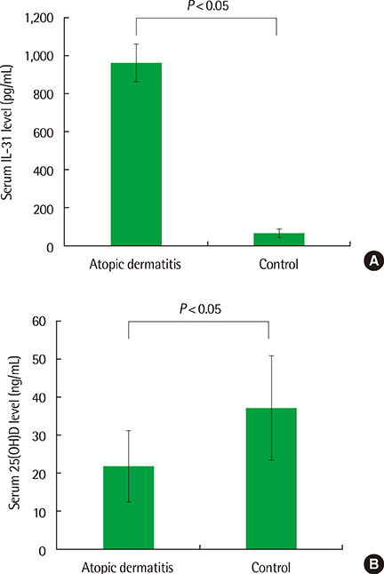
Fig. 1 Serum levels of 25(OH)D and IL-31 in children with atopic dermatitis and control group. (A) Serum IL-31 level was significantly higher in AD group compared to control group and (B) 25(OH)D level was significantly lower in AD group compared to control group. 25(OH)D, 25-hydroxyvitamin D; IL, interleukin; AD, atopic dermatitis.
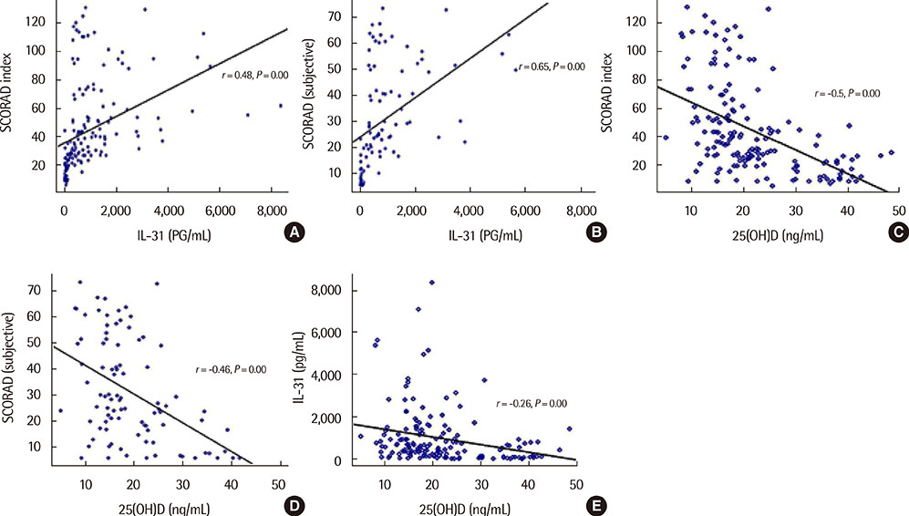
Fig. 2 Correlation between the SCORAD index, subjective SCORAD index, serum levels of 25(OH)D, and IL-31. The levels of IL-31 showed a positive correlation with the SCORAD index (A), and subjective SCORAD index (B). The levels of 25(OH)D were inversely correlated with the SCORAD index (C), and subjective SCORAD index (D). (E) The levels of 25(OH)D were inversely correlated with the IL-31 concentration. SCORAD, SCORing Atopic Dermatitis; 25(OH)D, 25-hydroxyvitamin D; IL, interleukin.
Cited by 2 articles
-
Current research status of pediatric atopic dermatitis in Korea
Bok Yang Pyun
Allergy Asthma Respir Dis. 2018;6(Suppl 1):S40-S43. doi: 10.4168/aard.2018.6.S1.S40.Relationship of serum vitamin D and interleukin-31 levels to allergic or nonallergic rhinitis in children
Seong Jun Park, Ji Eun Soh, Moon Soo Park, Hye Lim Jung, Jae Won Shim, Deok Soo Kim, Jung Yeon Shim
Allergy Asthma Respir Dis. 2018;6(1):41-46. doi: 10.4168/aard.2018.6.1.41.
Reference
-
1. Asher MI, Montefort S, Bjorksten B, Lai CK, Strachan DP, Weiland SK, et al. Worldwide time trends in the prevalence of symptoms of asthma, allergic rhinoconjunctivitis, and eczema in childhood: ISAAC Phases One and Three repeat multicountry cross-sectional surveys. Lancet. 2006; 368:733–743.
Article2. Stander S, Steinhoff M. Pathophysiology of pruritus in atopic dermatitis: an overview. Exp Dermatol. 2002; 11:12–24.
Article3. Holick MF. Vitamin D deficiency. N Engl J Med. 2007; 357:266–281.
Article4. Bikle D. Nonclassic actions of vitamin D. J Clin Endocrinol Metab. 2009; 94:26–34.
Article5. Grober U, Spitz J, Reichrath J, Kisters K, Holick MF. Vitamin D: Update 2013: From rickets prophylaxis to general preventive healthcare. Dermatoendocrinol. 2013; 5:331–347.6. Bonanno A, Gangemi S, La Grutta S, Malizia V, Riccobono L, Colombo P, et al. 25-Hydroxyvitamin D, IL-31, and IL-33 in children with allergic disease of the airways. Mediators Inflamm. 2014; 2014:520241.
Article7. Baumann R, Rabaszowski M, Stenin I, Gaertner-Akerboom M, Scheckenbach K, Wiltfang J, et al. The release of IL-31 and IL-13 after nasal allergen challenge and their relation to nasal symptoms. Clin Transl Allergy. 2012; 2:13.
Article8. Wittke A, Weaver V, Mahon BD, August A, Cantorna MT. Vitamin D receptor-deficient mice fail to develop experimental allergic asthma. J Immunol. 2004; 173:3432–3436.
Article9. Chiu YE, Havens PL, Siegel DH, Ali O, Wang T, Holland KE, et al. Serum 25-hydroxyvitamin D concentration does not correlate with atopic dermatitis severity. J Am Acad Dermatol. 2013; 69:40–46.
Article10. Zhang Q, Putheti P, Zhou Q, Liu Q, Gao W. Structures and biological functions of IL-31 and IL-31 receptors. Cytokine Growth Factor Rev. 2008; 19:347–356.
Article11. Dillon SR, Sprecher C, Hammond A, Bilsborough J, Rosenfeld-Franklin M, Presnell SR, et al. Interleukin 31, a cytokine produced by activated T cells, induces dermatitis in mice. Nat Immunol. 2004; 5:752–760.
Article12. Hanifin JM, Rajka G. Diagnostic features of atopic dermatitis. Acta Derm Venereol Suppl (Stockh). 1980; 92:44–47.13. Severity scoring of atopic dermatitis: the SCORAD index. Consensus Report of the European Task Force on Atopic Dermatitis. Dermatology. 1993; 186:23–31.14. Takaoka A, Arai I, Sugimoto M, Honma Y, Futaki N, Nakamura A, et al. Involvement of IL-31 on scratching behavior in NC/Nga mice with atopic-like dermatitis. Exp Dermatol. 2006; 15:161–167.
Article15. Sonkoly E, Muller A, Lauerma AI, Pivarcsi A, Soto H, Kemeny L, et al. IL-31: a new link between T cells and pruritus in atopic skin inflammation. J Allergy Clin Immunol. 2006; 117:411–417.
Article16. Kim S, Kim HJ, Yang HS, Kim E, Huh IS, Yang JM. IL-31 serum protein and tissue mRNA levels in patients with atopic dermatitis. Ann Dermatol. 2011; 23:468–473.
Article17. Raap U, Weibmantel S, Gehring M, Eisenberg AM, Kapp A, Folster-Holst R. IL-31 significantly correlates with disease activity and Th2 cytokine levels in children with atopic dermatitis. Pediatr Allergy Immunol. 2012; 23:285–288.
Article18. Ezzat MH, Hasan ZE, Shaheen KY. Serum measurement of interleukin-31 (IL-31) in paediatric atopic dermatitis: elevated levels correlate with severity scoring. J Eur Acad Dermatol Venereol. 2011; 25:334–339.
Article19. Palmer CN, Irvine AD, Terron-Kwiatkowski A, Zhao Y, Liao H, Lee SP, et al. Common loss-of-function variants of the epidermal barrier protein filaggrin are a major predisposing factor for atopic dermatitis. Nat Genet. 2006; 38:441–446.
Article20. Hata TR, Kotol P, Jackson M, Nguyen M, Paik A, Udall D, et al. Administration of oral vitamin D induces cathelicidin production in atopic individuals. J Allergy Clin Immunol. 2008; 122:829–831.
Article21. Samochocki Z, Bogaczewicz J, Jeziorkowska R, Sysa-Jedrzejowska A, Glinska O, Karczmarewicz E, et al. Vitamin D effects in atopic dermatitis. J Am Acad Dermatol. 2013; 69:238–244.
Article22. Camargo CA Jr, Ganmaa D, Sidbury R, Erdenedelger Kh, Radnaakhand N, Khandsuren B. Randomized trial of vitamin D supplementation for winter-related atopic dermatitis in children. J Allergy Clin Immunol. 2014; 134:831–835.e1.
Article23. Amestejani M, Salehi BS, Vasigh M, Sobhkhiz A, Karami M, Alinia H, et al. Vitamin D supplementation in the treatment of atopic dermatitis: a clinical trial study. J Drugs Dermatol. 2012; 11:327–330.24. Pichler J, Gerstmayr M, Szepfalusi Z, Urbanek R, Peterlik M, Willheim M. 1 alpha,25(OH)2D3 inhibits not only Th1 but also Th2 differentiation in human cord blood T cells. Pediatr Res. 2002; 52:12–18.
Article25. Boonstra A, Barrat FJ, Crain C, Heath VL, Savelkoul HF, O'Garra A. 1alpha,25-Dihydroxyvitamin d3 has a direct effect on naive CD4(+) T cells to enhance the development of Th2 cells. J Immunol. 2001; 167:4974–4980.
Article26. Jirapongsananuruk O, Melamed I, Leung DY. Additive immunosuppressive effects of 1,25-dihydroxyvitamin D3 and corticosteroids on TH1, but not TH2, responses. J Allergy Clin Immunol. 2000; 106:981–985.
Article27. Staeva-Vieira TP, Freedman LP. 1,25-dihydroxyvitamin D3 inhibits IFN-gamma and IL-4 levels during in vitro polarization of primary murine CD4+ T cells. J Immunol. 2002; 168:1181–1189.
Article28. Lemire JM, Archer DC, Beck L, Spiegelberg HL. Immunosuppressive actions of 1,25-dihydroxyvitamin D3: preferential inhibition of Th1 functions. J Nutr. 1995; 125:6 Suppl. 1704S–1708S.29. Hypponen E, Berry DJ, Wjst M, Power C. Serum 25-hydroxyvitamin D and IgE: a significant but nonlinear relationship. Allergy. 2009; 64:613–620.30. Cheon BR, Shin JE, Kim YJ, Shim JW, Kim DS, Jung HL, et al. Relationship between serum 25-hydroxyvitamin D and interleukin-31 levels, and the severity of atopic dermatitis in children. Korean J Pediatr. 2015; 58:96–101.
Article
- Full Text Links
-
- Actions
-
Cited
- CITED
-
- Close
- Share
-
- Similar articles
-
- Relationship between serum 25-hydroxyvitamin D and interleukin-31 levels, and the severity of atopic dermatitis in children
- Correlation between serum 25-hydroxyvitamin D levels and severity of atopic dermatitis in children
- Relationship of serum vitamin D and interleukin-31 levels to allergic or nonallergic rhinitis in children
- Vitamin D Status and Its Relationship with Disease Severity/activity in Patients with Atopic Dermatitis, Psoriasis, and Chronic Idiopathic Urticaria in Korea
- Correlation between serum 25-hydroxyvitamin D3 and the severity of atopic dermatitis in children with allergic or nonallergenic sensitization
