Corneal Biomechanical Properties of Normal Tension Glaucoma in Young Patients Evaluated with the Ocular Response Analyzer
- Affiliations
-
- 1Department of Ophthalmology, Ewha Womans University School of Medicine, Ewha Institute of Ophthalmology and Optometry, EIOO, Seoul, Korea. ckrey02@ewha.ac.kr
- KMID: 2216664
- DOI: http://doi.org/10.3341/jkos.2013.54.2.280
Abstract
- PURPOSE
To evaluate the corneal biomechanical properties and clinical characteristic of normal tension glaucoma (NTG) in young patients.
METHODS
We compared corneal biomechanical properties using an Ocular response analyzer (ORA) of under age 40 of 37 eyes of patients with NTG and 42 eyes of normal group.
RESULTS
The mean corneal resistance factor (CRF) and mean corneal hysteresis (CH) were significantly lower in NTG eyes (CRF, 9.2 +/- 2.1 mm Hg; CH, 9.8 +/- 1.8 mm Hg) than in normal eyes (CRF, 10.7 +/- 2.3 mm Hg; CH, 10.9 +/- 2.0 mm Hg; p = 0.01, p < 0.01). CH and CRF were associated with central corneal thickness (CCT) (CH; beta = 0.354, p < 0.01, CRF; beta = 0.348, p < 0.01) and glaucoma status (p < 0.01, p < 0.01).
CONCLUSIONS
The CRF and CH were significantly lower in NTG group while IOP cc was not significantly different between the group. In diagnosing the NTG in young patients, ORA maybe useful for distinguishing between the glaucoma eyes and normal eyes.
MeSH Terms
Figure
-
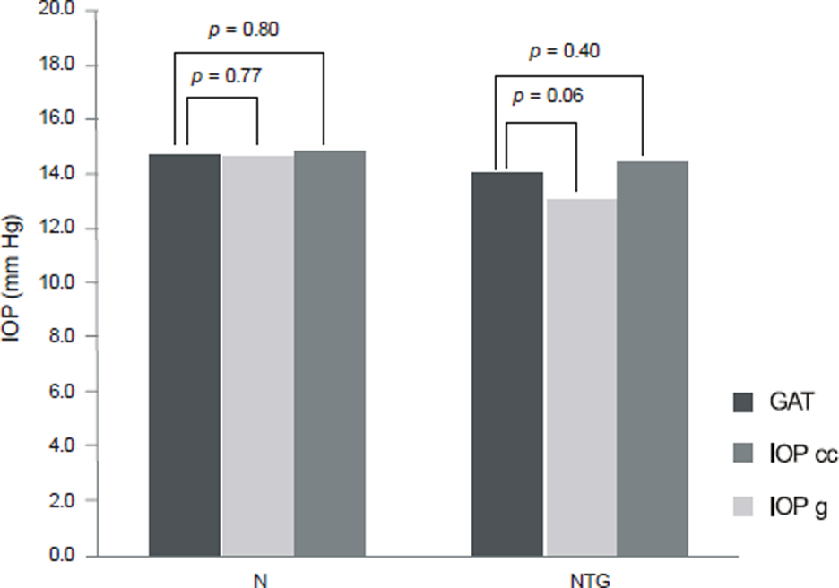
Figure 1. Intraocular pressure values of obtained with Ocular Response Analyzer and Goldman applanation tonometry in normal and normal tension glaucoma patient groups. N = Normal group; NTG = Normal tension glaucoma group; GAT = Goldmann applanation tonometer; IOP cc = cor-neal-compensated IOP; IOP g = Goldmann-correlated IOP.
Cited by 4 articles
-
The Effect of Corneal Biomechanical Factors on Ocular Pulse Amplitude in Normal Subjects
Han Jo Kwon, Ji Woong Lee, Jong Hun Shin
J Korean Ophthalmol Soc. 2015;56(8):1248-1255. doi: 10.3341/jkos.2015.56.8.1248.Diagnostic Availability of Ocular Response Analyzer in Korean Patients with Normal Tension Glaucoma
Ah Ran Cho, Yun Jeong Choi, Jin Young Rhew, Kyu Ryong Choi
J Korean Ophthalmol Soc. 2015;56(1):86-92. doi: 10.3341/jkos.2015.56.1.86.The Short-Term Effect of Prostaglandin Analog Monotherapy on Corneal Biomechanical Properties in Normal Tension Glaucoma Patients
Jung Yul Park, Ji Woong Lee, Jong Hoon Shin
J Korean Ophthalmol Soc. 2016;57(3):477-484. doi: 10.3341/jkos.2016.57.3.477.Diurnal Variation in Intraocular Pressure Measured by Ocular Response Analyzer in Korean Patients with Normal Tension Glaucoma
Yeon Jung Choi, Kyu Ryong Choi
J Korean Ophthalmol Soc. 2015;56(12):1913-1920. doi: 10.3341/jkos.2015.56.12.1913.
Reference
-
References
1. Shields MB. Normal-tension glaucoma: is it different from primary open-angle glaucoma? Curr Opin Ophthalmol. 2008; 19:85–8.
Article2. Drance S, Anderson DR, Schulzer M. Risk factors for progression of visual field abnormalities in normal-tension glaucoma. Am J Ophthalmol. 2001; 131:699–708.
Article3. Nakagami T, Yamazaki Y, Hayamizu F. Prognostic factors for progression of visual field damage in patients with normal-tension glaucoma. Jpn J Ophthalmol. 2006; 50:38–43.
Article4. Gordon MO, Beiser JA, Brandt JD, et al. The Ocular Hypertension Treatment Study: baseline factors that predict the onset of primary open-angle glaucoma. Arch Ophthalmol. 2002; 120:714–20.5. Herndon LW, Weizer JS, Stinnett SS. Central corneal thickness as a risk factor for advanced glaucoma damage. Arch Ophthalmol. 2004; 122:17–21.
Article6. Medeiros FA, Sample PA, Zangwill LM. Corneal thickness as a risk factor for visual field loss in patients with preperimetric glaucomatous optic neuropathy. Am J Ophthalmol. 2003; 136:805–13.
Article7. Pakravan M, Parsa A, Sanagou M, Parsa CF. Central corneal thickness and correlation to optic disc size: a potential link for susceptibility to glaucoma. Br J Ophthalmol. 2007; 91:26–8.
Article8. Argus WA. Ocular hypertension and central corneal thickness. Ophthalmology. 1995; 102:1810–2.
Article9. Copt RP, Thomas R, Mermoud A. Corneal thickness in ocular hypertension, primary open-angle glaucoma, and normal tension glaucoma. Arch Ophthalmol. 1999; 117:14–6.
Article10. Morad Y, Sharon E, Hefetz L, Nemet P. Corneal thickness and curvature in normal-tension glaucoma. Am J Ophthalmol. 1998; 125:164–8.
Article11. Bechmann M, Thiel MJ, Roesen B, et al. Central corneal thickness determined with optical coherence tomography in various types of glaucoma. Br J Ophthalmol. 2000; 84:1233–7.
Article12. Choi HJ, Kim DM, Hwang SS. Relationship between central cor-neal thickness and localized retinal nerve fiber layer defect in normal-tension glaucoma. J Glaucoma. 2006; 15:120–3.
Article13. Wu LL, Suzuki Y, Ideta R, Araie M. Central corneal thickness of normal tension glaucoma patients in Japan. Jpn J Ophthalmol. 2000; 44:643–7.
Article14. Iwase A, Suzuki Y, Araie M, et al. The prevalence of primary open-angle glaucoma in Japanese: the Tajimi Study. Ophthalmology. 2004; 111:1641–8.15. Tomidokoro A, Araie M, Iwase A. Tajimi Study Group. Corneal thickness and relating factors in a population-based study in Japan: the Tajimi Study. Am J Ophthalmol. 2007; 144:152–4.16. Xu L, Zhang H, Wang YX, Jonas JB. Central corneal thickness and glaucoma in adult Chinese: the Beijing Eye Study. J Glaucoma. 2008; 17:647–53.17. Luce DA. Determining in vivo biomechanical properties of the cornea with an ocular response analyzer. J Cataract Refract Surg. 2005; 31:156–62.
Article18. Congdon NG, Broman AT, Bandeen-Roche K, et al. Central cor-neal thickness and corneal hysteresis associated with glaucoma damage. Am J Ophthalmol. 2006; 141:868–75.
Article19. Bochmann F, Ang GS, Azuara-Blanco A. Lower corneal hysteresis in glaucoma patients with acquired pit of the optic nerve (APON). Graefes Arch Clin Exp Ophthalmol. 2008; 246:735–8.
Article20. Kirwan C, O'Keefe M, Lanigan B. Corneal hysteresis and intraocular pressure measurement in children using the reichert ocular response analyzer. Am J Ophthalmol. 2006; 142:990–2.
Article21. Sullivan-Mee M, Billingsley SC, Patel AD, et al. Ocular Response Analyzer in subjects with and without glaucoma. Optom Vis Sci. 2008; 85:463–70.
Article22. Wells AP, Garway-Heath DF, Poostchi A, et al. corneal hysteresis but not corneal thickness correlates with optic nerve surface compliance in glaucoma patients. Invest Ophthalmol Vis Sci. 2008; 49:3262–8.
Article23. Shah S, Laiquzzaman M, Mantry S, Cunliffe I. Ocular response analyser to assess hysteresis and corneal resistance factor in low tension, open angle glaucoma and ocular hypertension. Clin Experiment Ophthalmol. 2008; 36:508–13.
Article24. Ang GS, Bochmann F, Townend J, Azuara-Blanco A. Corneal bio-mechanical properties in primary open angle glaucoma and normal tension glaucoma. J Glaucoma. 2008; 17:259–62.
Article25. Abitbol O, Bouden J, Doan S, et al. Corneal hysteresis measured with the Ocular Response Analyzer in normal and glaucomatous eyes. Acta Ophthalmol. 2010; 88:116–9.26. Broman AT, Congdon NG, Bandeen-Roche K, Quigley HA. Influence of corneal structure, corneal responsiveness, and other ocular parameters on tonometric measurement of intraocular pressure. J Glaucoma. 2007; 16:581–8.
Article27. Burgoyne CF, Downs JC, Bellezza AJ, et al. The optic nerve head as a biomechanical structure: a new paradigm for understanding the role of IOP-related stress and strain in the pathophysiology of glaucomatous optic nerve head damage. Prog Retin Eye Res. 2005; 24:39–73.
Article28. Lesk MR, Hafez AS, Descovich D. Relationship between central corneal thickness and changes of optic nerve head topography and blood flow after intraocular pressure reduction in open-angle glaucoma and ocular hypertension. Arch Ophthalmol. 2006; 124:1568–72.
Article29. Edmund C. Corneal elasticity and ocular rigidity in normal and keratoconic eyes. Acta Ophthalmol. 1988; 66:134–40.
Article30. McBrien NA, Gentle A. Role of the sclera in the development and pathological complications of myopia. Prog Retin Eye Res. 2003; 22:307–38.
Article31. Miller KM, Quigley HA. Comparison of optic disc features in low-tension and typical open-angle glaucoma. Ophthalmic Surg. 1987; 18:882–9.
Article32. Javitt JC, Spaeth GL, Katz LJ, et al. Acquired pits of the optic nerve. Increased prevalence in patients with low-tension glaucoma. Ophthalmology. 1990; 97:1038–43.33. Johnson CS, Mian SI, Moroi S, et al. Role of corneal elasticity in damping of intraocular pressure. Invest Ophthalmol Vis Sci. 2007; 48:2540–4.
Article34. Downs JC, Suh JK, Thomas KA, et al. Viscoelastic material properties of the peripapillary sclera in normal and early-glaucoma monkey eyes. Invest Ophthalmol Vis Sci. 2005; 46:540–6.
Article35. Sigal IA, Flanagan JG, Ethier CR. Factors influencing optic nerve head biomechanics. Invest Ophthalmol Vis Sci. 2005; 46:4189–99.
Article36. Shah S, Laiquzzaman M, Cunliffe I, Mantry S. The use of the Reichert ocular response analyser to establish the relationship between ocular hysteresis, corneal resistance factor and central cor-neal thickness in normal eyes. Cont Lens Anterior Eye. 2006; 29:257–62.
Article37. Pepose JS, Feigenbaum SK, Qazi MA, et al. Changes in corneal biomechanics and intraocular pressure following LASIK using static, dynamic, and noncontact tonometry. Am J Ophthalmol. 2007; 143:39–47.
Article38. Hager A, Loge K, Schroeder B, et al. Effect of central corneal thickness and corneal hysteresis on tonometry as measured by dynamic contour tonometry, ocular response analyzer, and Goldmann tonometry in glaucomatous eyes. J Glaucoma. 2008; 17:361–5.39. Lam A, Chen D, Chiu R, Chui WS. Comparison of IOP measurements between ORA and GAT in normal Chinese. Optom Vis Sci. 2007; 84:909–14.
Article40. Casson RJ, Abraham LM, Newland HS, et al. Corneal thickness and intraocular pressure in a nonglaucomatous Burmese population: the Meiktila Eye Study. Arch Ophthalmol. 2008; 126:981–5.41. Radhakrishnan H, Miranda MA, O'Donnell C. Corneal bio-mechanical properties and their correlates with refractive error. Clin Exp Optom. 2012; 95:12–8.
Article42. Jiang Z, Shen M, Mao G, et al. Association between corneal bio-mechanical properties and myopia in Chinese subjects. Eye. 2011; 25:1083–9.
Article43. Chang PY, Chang SW, Wang JY. Assessment of corneal bio-mechanical properties and intraocular pressure with the Ocular Response Analyzer in childhood myopia. Br J Ophthalmol. 2010; 94:877–81.
Article44. Song Y, Congdon N, Li L, et al. Corneal hysteresis and axial length among Chinese secondary school children: the Xichang Pediatric Refractive Error Study (X-PRES)report no. (4):Am J Ophthalmol. 2008; 145:819–26.45. Lim L, Gazzard G, Chan YH, et al. Cornea biomechanical characteristics and their correlates with refractive error in Singaporean children. Invest Ophthalmol Vis Sci. 2008; 49:3852–7.
Article46. Shen M, Wang J, Qu J, et al. Diurnal variation of ocular hysteresis, corneal thickness, and intraocular pressure. Optom Vis Sci. 2008; 85:1185–92.
Article
- Full Text Links
-
- Actions
-
Cited
- CITED
-
- Close
- Share
-
- Similar articles
-
- The Association Between Corneal Biomechanical Properties and Progression of Visual Field Loss in Normal Tension Glaucoma
- The Short-Term Effect of Prostaglandin Analog Monotherapy on Corneal Biomechanical Properties in Normal Tension Glaucoma Patients
- The Association between Corneal Biomechanical Properties and Initial Visual Field Defect Pattern in Normal Tension Glaucoma
- Diurnal Variation in Intraocular Pressure Measured by Ocular Response Analyzer in Korean Patients with Normal Tension Glaucoma
- Diagnostic Availability of Ocular Response Analyzer in Korean Patients with Normal Tension Glaucoma
