J Korean Ophthalmol Soc.
2011 Jun;52(6):658-664. 10.3341/jkos.2011.52.6.658.
Analysis of CCL5 Concentration in Tears of Dry Eye Patients
- Affiliations
-
- 1Department of Ophthalmology, Chonnam National University Hospital, Chonnam National University Medical School, Gwangju, Korea. kcyoon@chonnam.ac.kr
- KMID: 2214636
- DOI: http://doi.org/10.3341/jkos.2011.52.6.658
Abstract
- PURPOSE
To investigate the expression of CCL5/RANTES (regulated upon activation, normal T cell expressed and secreted) in the tears of dry eye patients.
METHODS
Forty patients with dry eye (15 Sjogren's and 25 non-Sjogren's syndrome patients) and ten control subjects were recruited for the present study. The concentration of RANTES in tears was measured using an enzyme-linked immunosorbent assay. The correlations between RANTES level, tear film and ocular surface parameters, including tear film break-up time, basal tear secretion, tear clearance rate, corneal sensation, keratoepitheliopathy, and conjunctival goblet cell density, were analyzed in patients with dry eye syndrome.
RESULTS
The concentrations of RANTES were 435.46 +/- 104.45 pg/ml in Sjogren's syndrome patients, 257.42 +/- 46.72 pg/ml in non-Sjogren's syndrome patients, and 97.53 +/- 29.15 pg/ml in the control patients (p < 0.01). The levels correlated significantly with basal tear secretion, tear clearance rate, keratoepitheliopathy, and goblet cell density (p < 0.05).
CONCLUSIONS
CCL5/RANTES level increases in the tears of dry eye patients and correlates with various tear film and ocular surface parameters.
Keyword
MeSH Terms
Figure
-

Figure 1. RANTES levels in tears of Sjögren's syndrome patients, non-Sjögren's syndrome patients and control subjects. SS = Sjögren's syndrome. * p < 0.05.
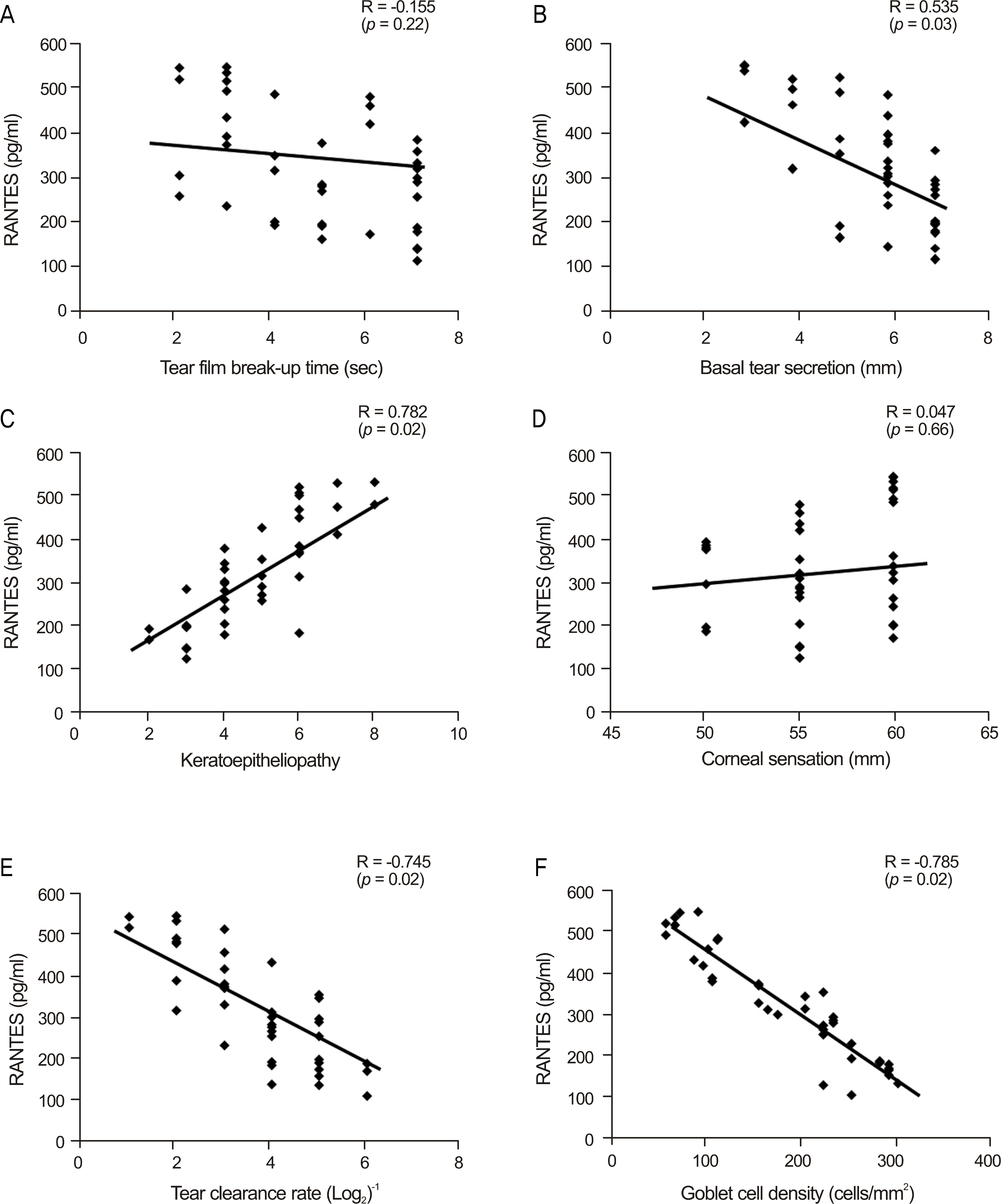
Figure 2. Correlation between RANTES levels in tears of dry eye patients and tear surface parameters including tear film break-up time (A), basal tear secretion (B), keratoepitheliopathy score (C), corneal sensation (D), tear clearance rate (E), and conjunctival goblet cell density (F).
Cited by 1 articles
-
Clinical Features of Dry Eye in Thyroid-Associated Ophthalmopathy According to Disease Activity
Jun Young Ha, Won Choi, Kyung Chul Yoon
J Korean Ophthalmol Soc. 2016;57(7):1037-1043. doi: 10.3341/jkos.2016.57.7.1037.
Reference
-
References
1. The definition and classification of dry eye disease: report of the Definition and Classification Subcommittee of the International Dry Eye WorkShop (2007). Ocul Surf. 2007; 5:75–92.2. Moss SE, Klein R, Klein BE. Prevalence of and risk factors for dry eye syndrome. Arch Ophthalmol. 2000; 118:1264–8.
Article3. Lee AJ, Lee J, Saw SM, et al. Prevalence and risk factors associated with dry eye symptoms: a population based study in Indonesia. Br J Ophthalmol. 2002; 86:1347–51.
Article4. Uchino M, Uchino Y, Dogru M, et al. Dry eye disease in Japan: An epidemiologic study. Cornea. 2009; 28:S31–4.
Article5. Stern ME, Gao J, Schwalb TA, et al. Conjunctival T-cell sub-populations in Sjögren's and non-Sjögren's patients with dry eye. Invest Ophthalmol Vis Sci. 2002; 43:2609–14.6. Rolando M, Barabino S, Mingari C, et al. Distribution of conjunctival HLA-DR expression and the pathogenesis of damage in early dry eyes. Cornea. 2005; 24:951–4.
Article7. Pflugfelder SC, Jones D, Ji Z, et al. Altered cytokine balance in the tear fluid and conjunctiva of patients with Sjögren's syndrome keratoconjunctivitis sicca. Curr Eye Res. 1999; 19:201–11.
Article8. Yoon KC, Jeong IY, Park YG, Yang SY. Interleukin-6 and tumor necrosis factor-alpha levels in tears of patients with dry eye syndrome. Cornea. 2007; 26:431–7.9. Corrales RM, Stern ME, De Paiva CS, et al. Desiccating stress stimulates expression of matrix metalloproteinases by the corneal epithelium. Invest Ophthalmol Vis Sci. 2006; 47:3293–302.
Article10. Baudouin C, Liang H, Bremond-Gignac D, et al. CCR 4 and CCR 5 expression in conjunctival specimens as differential markers of T(H)1/ T(H)2 in ocular surface disorders. J Allergy Clin Immunol. 2005; 116:614–9.11. Gulati A, Sacchetti M, Bonini S, Dana R. Chemokine receptor CCR5 expression in conjunctival epithelium of patients with dry eye syndrome. Arch Ophthalmol. 2006; 124:710–6.
Article12. Yoon KC, De Paiva CS, Qi H, et al. Expression of Th-1 chemo-kines and chemokine receptors on the ocular surface of C57BL/6 mice: effects of desiccating stress. Invest Ophthalmol Vis Sci. 2007; 48:2561–9.
Article13. Yoon KC, De Paiva CS, Qi H, et al. Desiccating environmental stress exacerbates autoimmune lacrimal keratoconjunctivitis in non-obese diabetic mice. J Autoimmun. 2008; 30:212–21.
Article14. Yoon KC, Park CS, You IC, et al. Expression of CXCL9, −10, −11, and CXCR3 in the tear film and ocular surface of patients with dry eye syndrome. Invest Ophthalmol Vis Sci. 2010; 51:643–50.
Article15. Kim HG, You IC, Yoon KC. I-TAC concentration in tears of dry eye patients and its correlation with tear surface parameters. J Korean Ophthalmol Soc. 2008; 49:1565–71.
Article16. Vitali C, Bombardieri S, Jonsson R, et al. Classification criteria for Sjögren's syndrome: a revised version of the European criteria pro-posed by the American-European Consensus Group. Ann Rheum Dis. 2002; 61:554–8.17. Dogru M, Katakami C, Inoue M. Tear function and ocular surface changes in noninsulin-dependent diabetes mellitus. Ophthalmology. 2001; 108:586–92.
Article18. Yoon KC, Jeong IY, Park YG, Yang SY. Interleukin-6 and tumor necrosis factor-alpha levels in tears of patients with dry eye syndrome. Cornea. 2007; 26:431–7.19. Pflugfelder SC. Anti-inflammatory therapy of dry eye. Ocul Surf. 2003; 1:31–6.
Article20. Jones DT, Monroy D, Ji Z, et al. Sjögren's syndrome: cytokine and Epstein-Barr viral gene expression within the conjunctival epithelium. Invest Ophthalmol Vis Sci. 1994; 35:3493–504.21. Lee SH, Im SK, Yoon KC. CCL4 concentration in tears of dry eye patients and its correlation with tear surface parameters. J Korean Ophthalmol Soc. 2010; 51:313–9.
Article22. Malesiń ski R, Bakunowicz-Ł azarczyk A, Wysocka J. The role of chemokines CCL3/ MIP-1 alfa and CCL4/ MIP-1 beta in pathogenesis of dry eye syndrome. Klin Oczna. 2008; 110:277–9.23. Ogawa N, Kawanami T, Shimoyama K, et al. Expression of interferon-inducible T cell alpha chemoattractant (CXCL11) in the sali-vary glands of patients with Sjögren's syndrome. Clin Immunol. 2004; 112:235–8.24. Petkovic V, Moghini C, Paoletti S, et al. I-TAC/CXCL11 is a natu-ral antagonist for CCR5. J Leukoc Biol. 2004; 76:701–8.
Article25. Donlon TA, Krensky AM, Wallace MR, et al. Localization of a human T-cell-specific gene, RANTES (D17S136E), to chromosome 17q11.2-q12. Genomics. 1990; 6:548–53.
Article26. Maghazachi AA, Al-Aoukaty A, Schall TJ. CC chemokines induce the generation of killer cells from CD56+ cells. Eur J Immunol. 1996; 26:315–9.
Article27. Song A, Nikolcheva T, Krensky AM. Transcriptional regulation of RANTES expression in T lymphocytes. Immunol Rev. 2000; 177:236–45.28. Cocchi F, DeVico AL, Garzino-Demo A, et al. Identification of RANTES, MIP-1 alpha, and MIP-1 beta as the major HIV-sup-pressive factors produced by CD8+ T cells. Science. 1995; 270:1811–5.29. Vangelista L, Secchi M, Liu X, et al. Engineering of Lactobacillus jensenii to secrete RANTES and a CCR5 antagonist analogue as live HIV-1 blockers. Antimicrob Agents Chemother. 2010; 54:2994–3001.30. Bacon KB, Premack BA, Gardner P, Schall TJ. Activation of dual T cell signaling pathways by the chemokine RANTES. Science. 1995; 269:1727–30.
Article31. Appay V, Dunbar PR, Cerundolo V, et al. RANTES activates anti-gen-specific cytotoxic T lymphocytes in a mitogen-like manner through cell surface aggregation. Int Immunol. 2000; 12:1173–82.
Article32. Turner L, Ward SG, Westwick J. RANTES-activated human T lymphocytes. A role for phosphoinositide 3-kinase. J Immunol. 1995; 155:2437–44.
- Full Text Links
-
- Actions
-
Cited
- CITED
-
- Close
- Share
-
- Similar articles
-
- The Concentration of Tear Lysozyme in Normal Subjects and Dry Eye
- Recent treatment of dry eye
- CCL4 Concentration in Tears of Dry Eye Patients and Its Correlation With Tear Surface Parameters
- The significance of tear film break-up time in the diagnosis of dry eye syndrome
- Changes in Total Tear Protein and Lipocalin Concentration According to Frequency of Artificial Tear Usage
