Papilledema Associated with Anemia in a Patient with Paroxysmal Nocturnal Hemoglobinuria
- Affiliations
-
- 1Department of Ophthalmology, Chungbuk National University College of Medicine, Cheongju, Korea. simple521@chungbuk.ac.kr
- KMID: 2211002
- DOI: http://doi.org/10.3341/jkos.2007.48.10.1438
Abstract
-
PURPOSE: To report a case of bilateral optic disc edema associated with hemolytic anemia and paroxysmal nocturnal hemoglobinuria (PNH).
METHODS
A 51-year-old woman visited our ophthalmologic clinic complaining of metamorphopsia. Twenty eight years ago, she had been diagnosed with PNH and hemolytic anemia and had received blood transfusion on an irregular basis. The best corrected visual acuity was initially 0.5 in the right eye and 1.0 in the left eye. Light reflex was intact and no afferent pupillary defect was found. Fundus examination revealed severe optic disc swellings with indistinct margins in both eyes. Papillary and peripapillary retinal hemorrhages were also present.
RESULTS
A visual field test revealed the enlarged physiologic scotoma in both eyes. Fluorescein angiograms showed hyperfluorescence of the optic disc and blocked fluorescence due to the papillary hemorrhages. Optical coherence tomograms of the optic disc showed the loss of physiologic disc cupping and severe elevation. There was no evidence of an intracranial lesion upon brain magnetic resonance imaging. These findings were compatible with optic disc edema associated with anemia and the management was oriented towards the anemia. At the 2-months follow-up, the best corrected visual acuity of both eyes had improved to 1.0 and optic disc edema markedly decreased. However, the patient's overall physical condition deteriorated and she expired due to dyspnea and hepatic coma.
MeSH Terms
-
Anemia*
Anemia, Hemolytic
Blood Transfusion
Brain
Dyspnea
Edema
Female
Fluorescein
Fluorescence
Follow-Up Studies
Hemoglobinuria, Paroxysmal*
Hemorrhage
Hepatic Encephalopathy
Humans
Magnetic Resonance Imaging
Middle Aged
Papilledema*
Pupil Disorders
Reflex
Retinal Hemorrhage
Scotoma
Vision Disorders
Visual Acuity
Visual Field Tests
Fluorescein
Figure
-
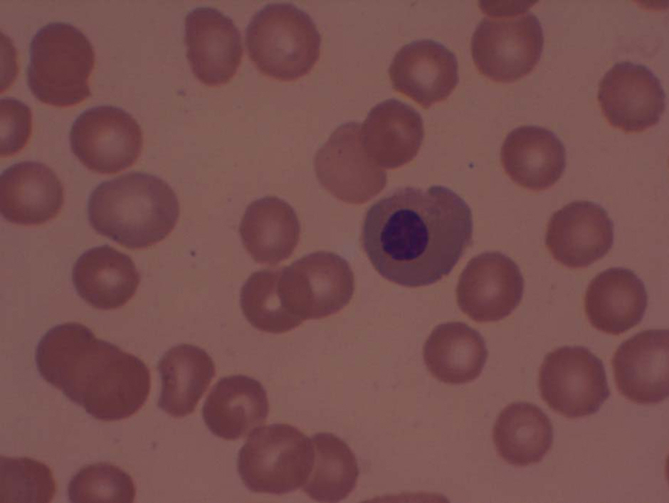
Figure 1. Photograph of peripheral blood smear. The erythrocytes are relatively normal in size and in hemoglobin content, but they are insufficient in number. These findings correspond to normocytic normochromic features of hemolytic anemia (Wright-Giemsa stain, χ400).
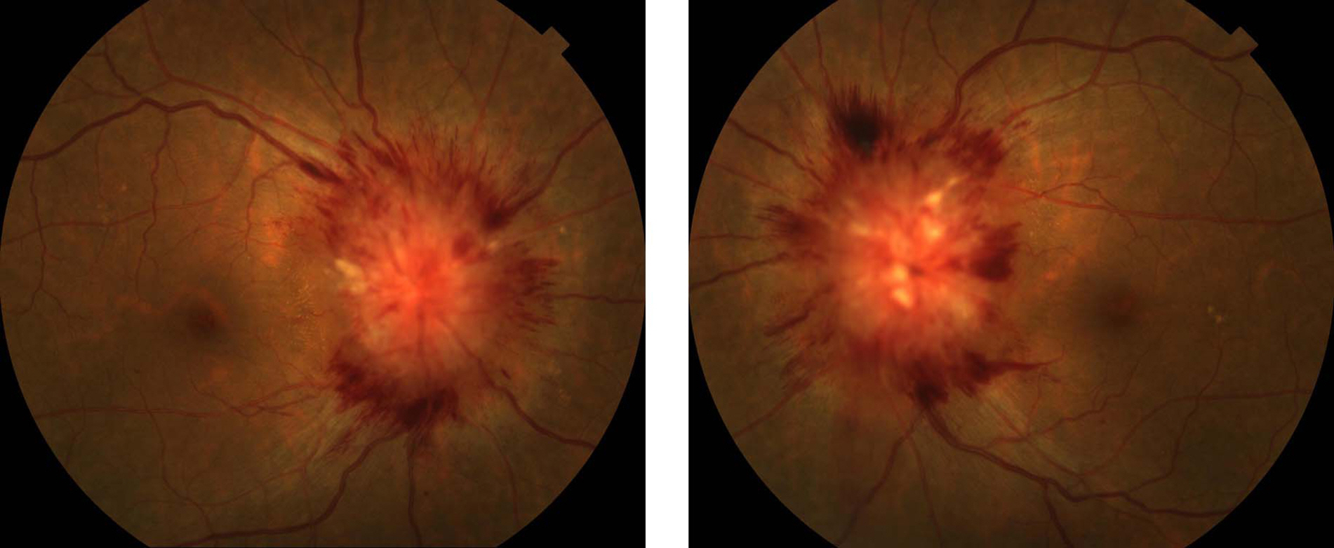
Figure 2. Fundus photographs show bilateral disc edema at the first visit. The disc margin is indistinct and papillary and peripapillary retinal hemorrhages are accompanied.
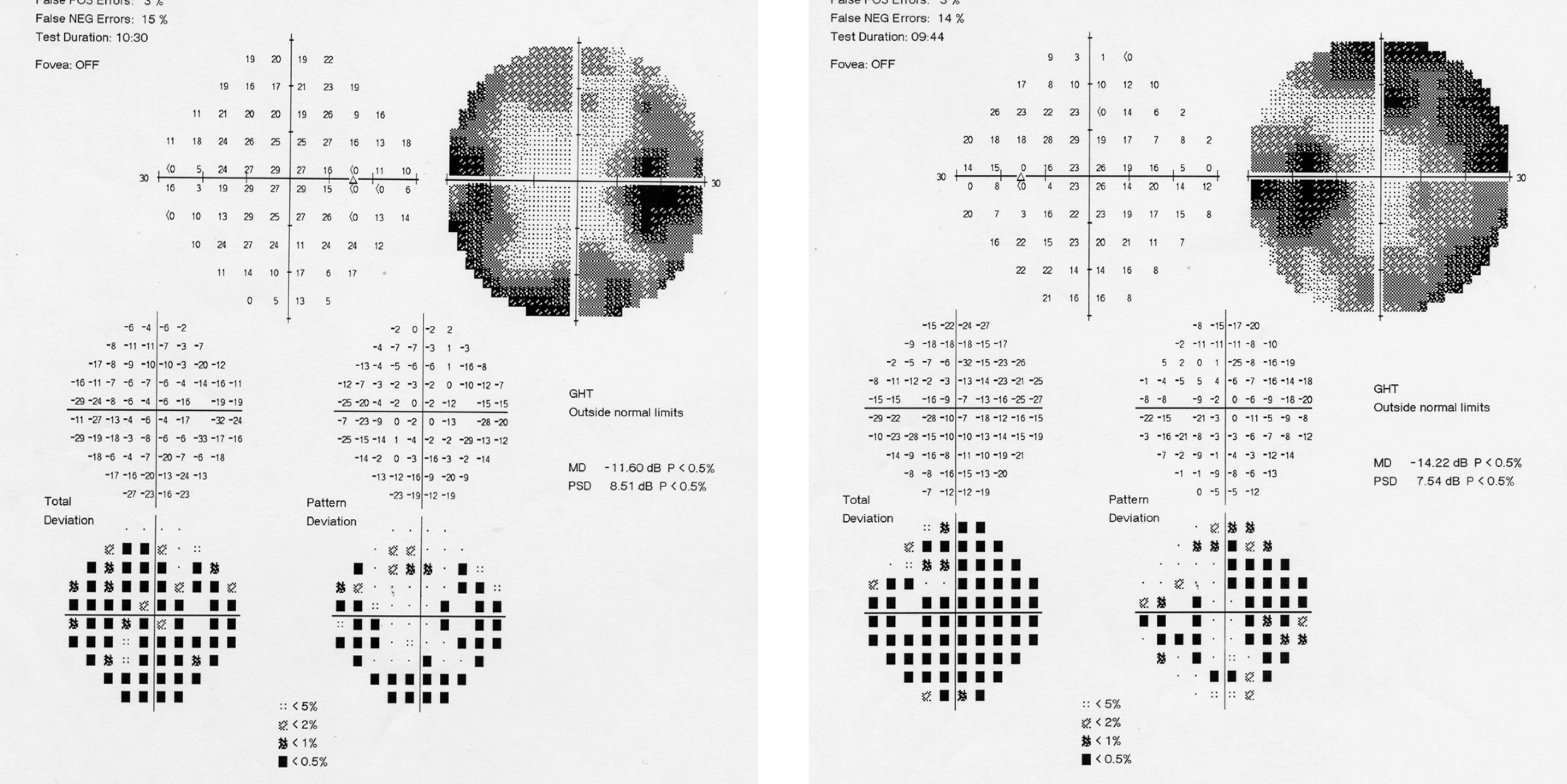
Figure 3. Automated Humphrey visual field test demonstrates enlarged physiologic scotoma and irregular peripheral visual field defects.
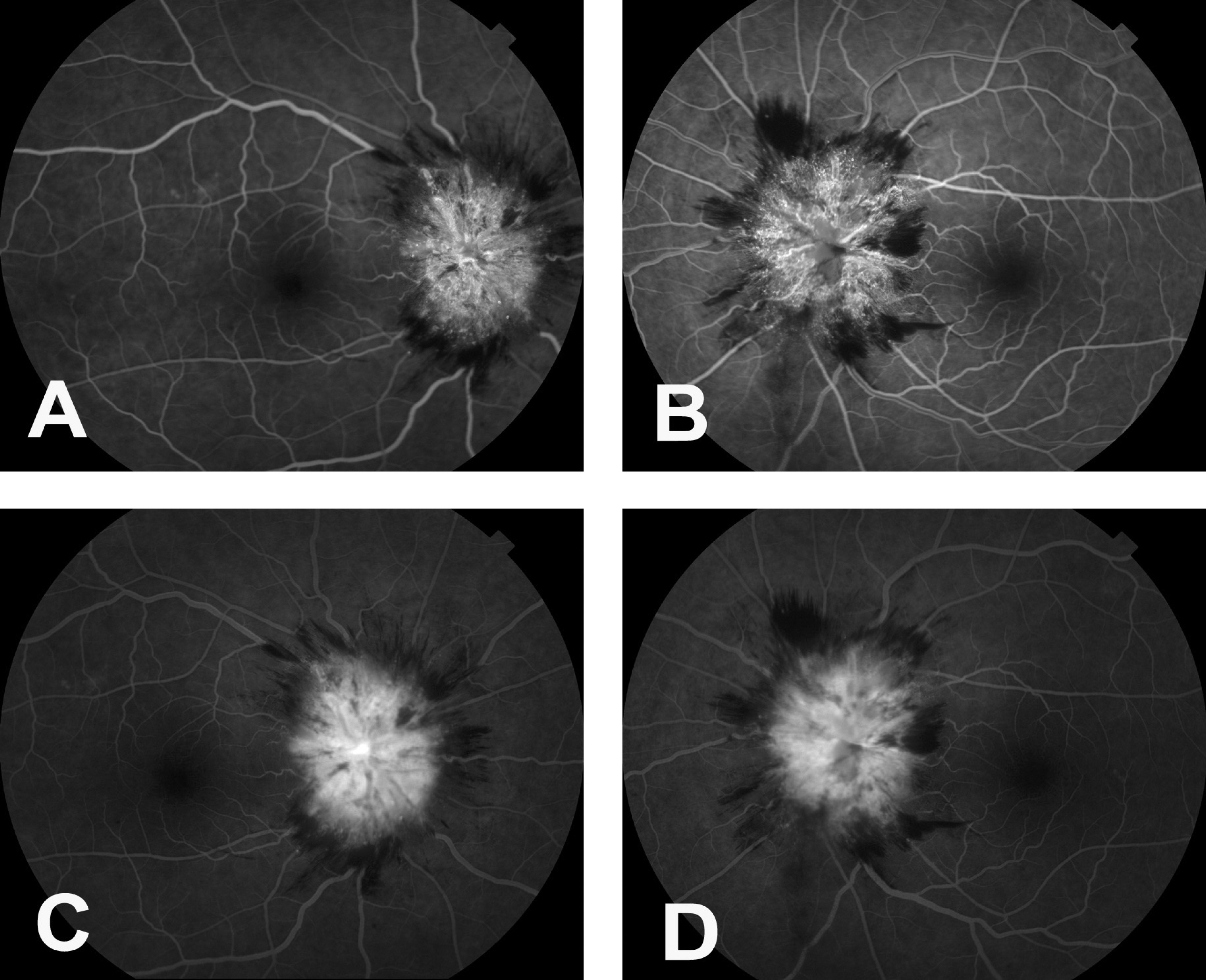
Figure 4. Early (A, B) and late (C, D) phase fluorescein angiograms show continued hyperfluorescence of the disc accompanied blocked fluorescence by papillary and peripapillary hemorrhages.

Figure 5. Optical coherence tomographs of the right (A) and left (B) optic disc show the loss of physiologic disc cupping and elevated optic disc.
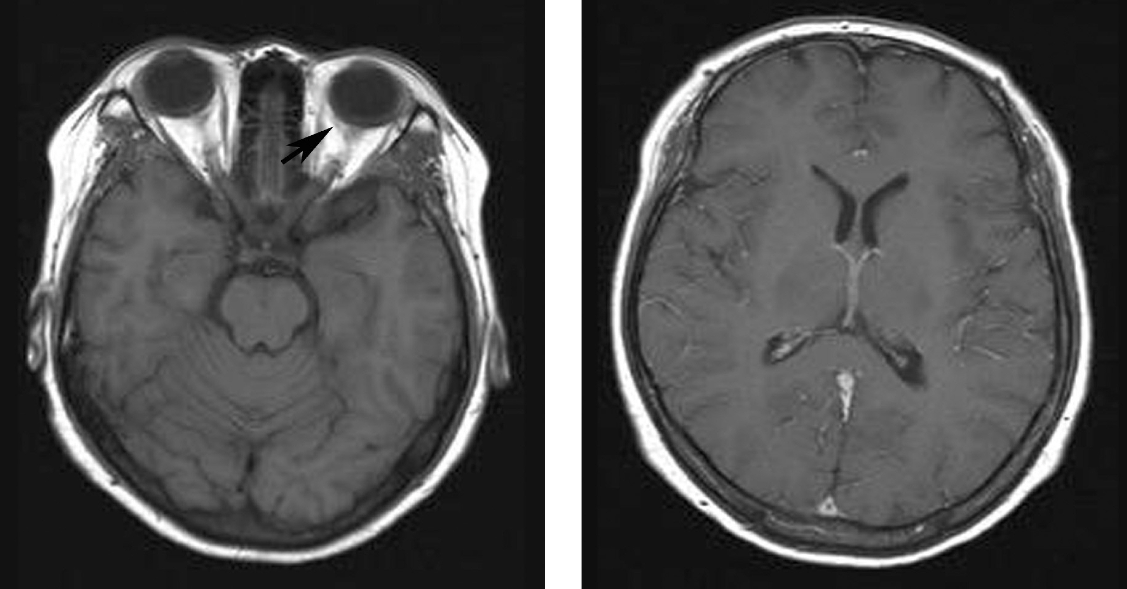
Figure 6. Orbit (A) and brain (B) MRIs of the patient reveal no evidence of retrobulbar lesion and brain metastasis or hydrocephalus. The superior ophthalmic vein (arrow) is not dilated.
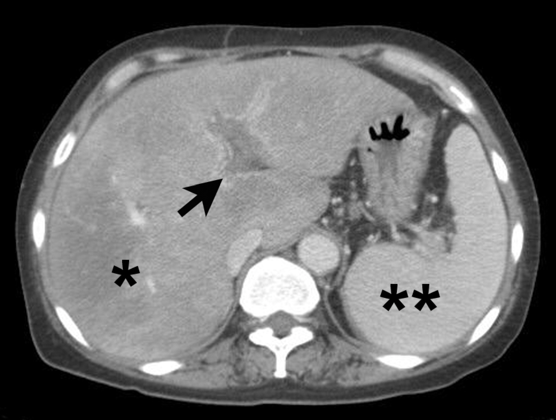
Figure 7. Abdomen CT shows diffuse swelling of the liver (*) and spleen (**) with left portal vein thrombosis (arrow).
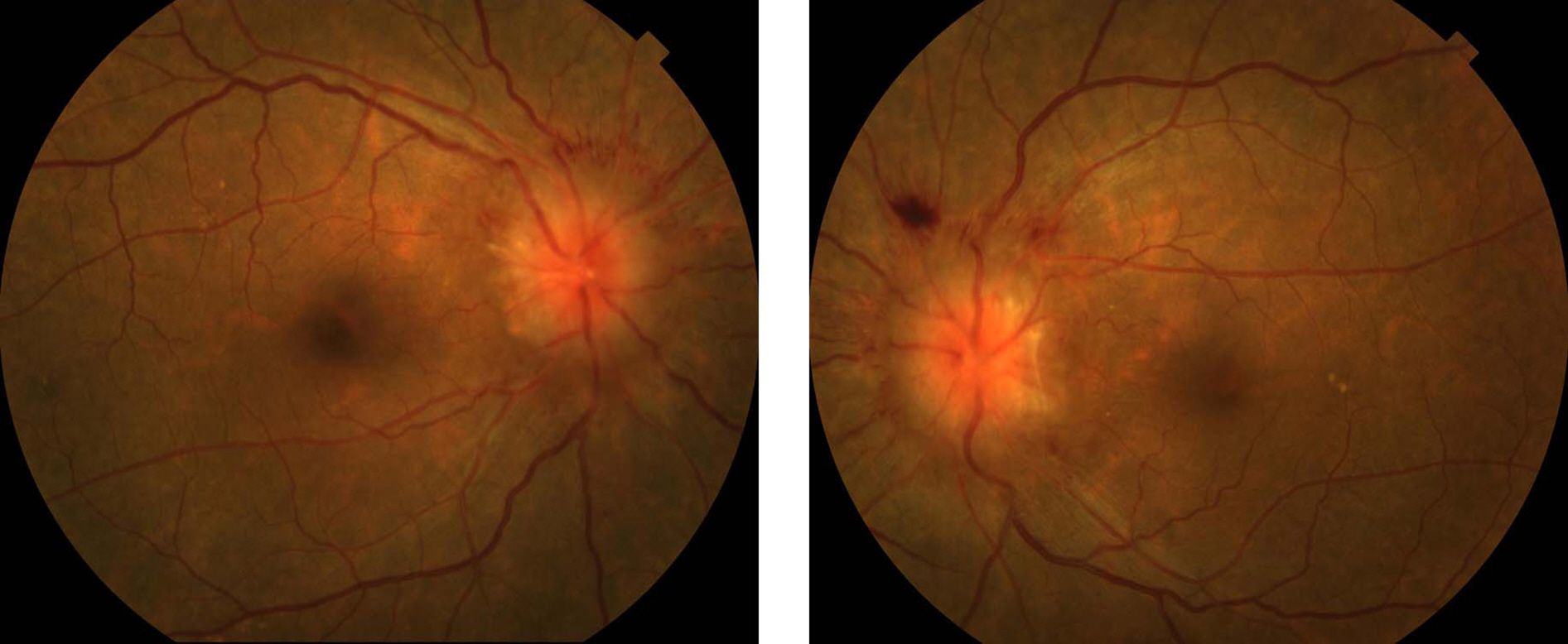
Figure 8. Fundus photographs after 2 months show improved papilledema.
Reference
-
References
1. Rosse WF, Parker CJ. Paroxysmal nocturnal haemoglobinuria. Clin Haematol. 1985; 14:105–25.
Article2. Rosse WF. Phosphatidylinositol-linked proteins and paroxysmal nocturnal hemoglobinuria. Blood. 1990; 75:1595–601.
Article3. Rotoli B, Luzzatto L. Paroxysmal nocturnal hemoglobinuria. Semin Hematol. 1989; 26:201–7.4. Song JK, Yoon ZW. Fundus findings in leukemia and various anemias. J Korean Ophthalmol Soc. 1975; 16:124–128.5. Lubeck NJ. Papilledema caused by iron deficiency anemia. Trans Am Acad Ophthalmol Otolaryngol. 1959; 63:306–10.6. Schwaber JR, Blumberg AG. Papilledema associated with blood loss anemia. Ann Intern Med. 1961; 55:1004–7.
Article7. Ikkala E, Laitinen L. Papilloedema due to iron deficiency anemia. Acta Haematol. 1963; 29:368–70.8. Stoebner R, Kiser R, Alperin JB. Iron deficiency anemia and papilledema. Rapid resolution with oral iron therapy. Am J Dig Dis. 1970; 15:919–22.9. Lilley ER, Bruggers CS, Pollock SC. Papilledema in a patient with aplastic anemia. Arch Ophthalmol. 1990; 108:1674–5.
Article10. Saleh T, Green W. Bilateral reversible optic disc oedema associated with iron deficiency anemia. Eye. 2000; 14:672–3.11. Taylor JP, Galetta SL, Asbury AK, Volpe NJ. Hemolytic anemia presenting as idiopathic intracranial hypertension. Neurology. 2002; 59:960–1.
Article12. Nazir SA, Siatkowski RM. Pseudotumor cerebri in idiopathic aplastic anemia. J AAPOS. 2003; 7:71–4.
Article13. Biousse V, Rucker JC, Vignal C, et al. Anemia and papilledema. Am J Ophthalmol. 2003; 135:437–46.
Article14. Aktan S, Kansu T, Kansu E, Zileli T. Papilledema in paroxysmal nocturnal hemoglobinuria. J Clin Neuroophthalmol. 1984; 4:47–8.15. al-Hakim M, Katirji B, Osorio I, Weisman R. Cerebral venous thrombosis in paroxysmal nocturnal hemoglobinuria: report of two cases. Neurology. 1993; 43:742–6.
Article16. Hauser D, Barzilai N, Zalish M, et al. Bilateral papilledema with retinal hemorrhages in association with cerebral venous sinus thrombosis and paroxysmal nocturnal hemoglobinuria. Am J Ophthalmol. 1996; 122:592–3.
Article17. Capriles L. Intracranial hypertension and iron deficiency anemia. Arch Neurol. 1963; 9:147–53.18. Trujillo MH, Desenne JJ, Pinto HB. Reversible papilledema in iron deficiency anemia. Ann Ophthalmol. 1972; 4:378–80.19. Foester HS. Optic disc edema due to iron deficiency. Occurrence with normal cerebrospinal fluid pressure. Conn Med. 1985; 49:290–2.20. Zimmerman D, Bell WR. Venous thrombosis and splenic rupture in paroxysmal nocturnal hemoglobinuria. Am J Med. 1980; 68:275–9.
Article21. Wiedmer T, Hall SE, Ortel TL, et al. Complement-induced vesiculation and exposure of membrane prothrombinase sites in platelets of paroxysmal nocturnal hemoglobinuria. Blood. 1993; 82:1192–6.
Article22. Walker RW. Idiopathic intracranial hypertension: any light on the mechanism of the raised pressure? J Neurol Neurosurg Psychiatry. 2001; 71:1–5.
Article23. Kim IT, Chang SD. Papilledema and cerebral venous thrombosis in a patient with systemic lupus erythematosis. J Korean Ophthalmol Soc. 1999; 40:2015–9.24. Capriles LF. Intracranial hypertension and iron deficiency anemia: report of four cases. Arch Neurol. 1963; 9:147–53.
- Full Text Links
-
- Actions
-
Cited
- CITED
-
- Close
- Share
-
- Similar articles
-
- A Case of Cerebral Venous Thrombosis in Paroxysmal Nocturnal Hemoglobinuria
- Paroxysmal Nocturnal Hemoglobinuria
- Paroxysmal Nocturnal Hemoglobinuria Presenting as Recurrent Jejunitis
- Delivery in a Patient with Paroxysmal Nocturnal Hemoglobinuria Successfully Managed with Low Molecular Weight Heparin Therapy
- Paroxysmal Nocturnal Hemoglobinuria Presenting with Chronic Abdominal Pain and Iron Deficiency Anemia
