J Korean Soc Spine Surg.
2012 Jun;19(2):72-76. 10.4184/jkss.2012.19.2.72.
Ossified Meningioma: A Case Report
- Affiliations
-
- 1Department of Orthopedic Surgery, Seoul Sacred Heart General Hospital, Seoul, Korea. msh124@paran.com
- KMID: 2209560
- DOI: http://doi.org/10.4184/jkss.2012.19.2.72
Abstract
- STUDY DESIGN: A case report
OBJECTIVES
To report an extremely rare case of the spinal meningioma containing bone. SUMMARY OF LITERATURE REVIEW: Spinal meningiomas represent 16.6-46.7% of the primary spinal tumors and 1 to 5% of them are calcified. Ossification is an extremely uncommon event that complicates the resection surgery.
MATERIALS AND METHODS
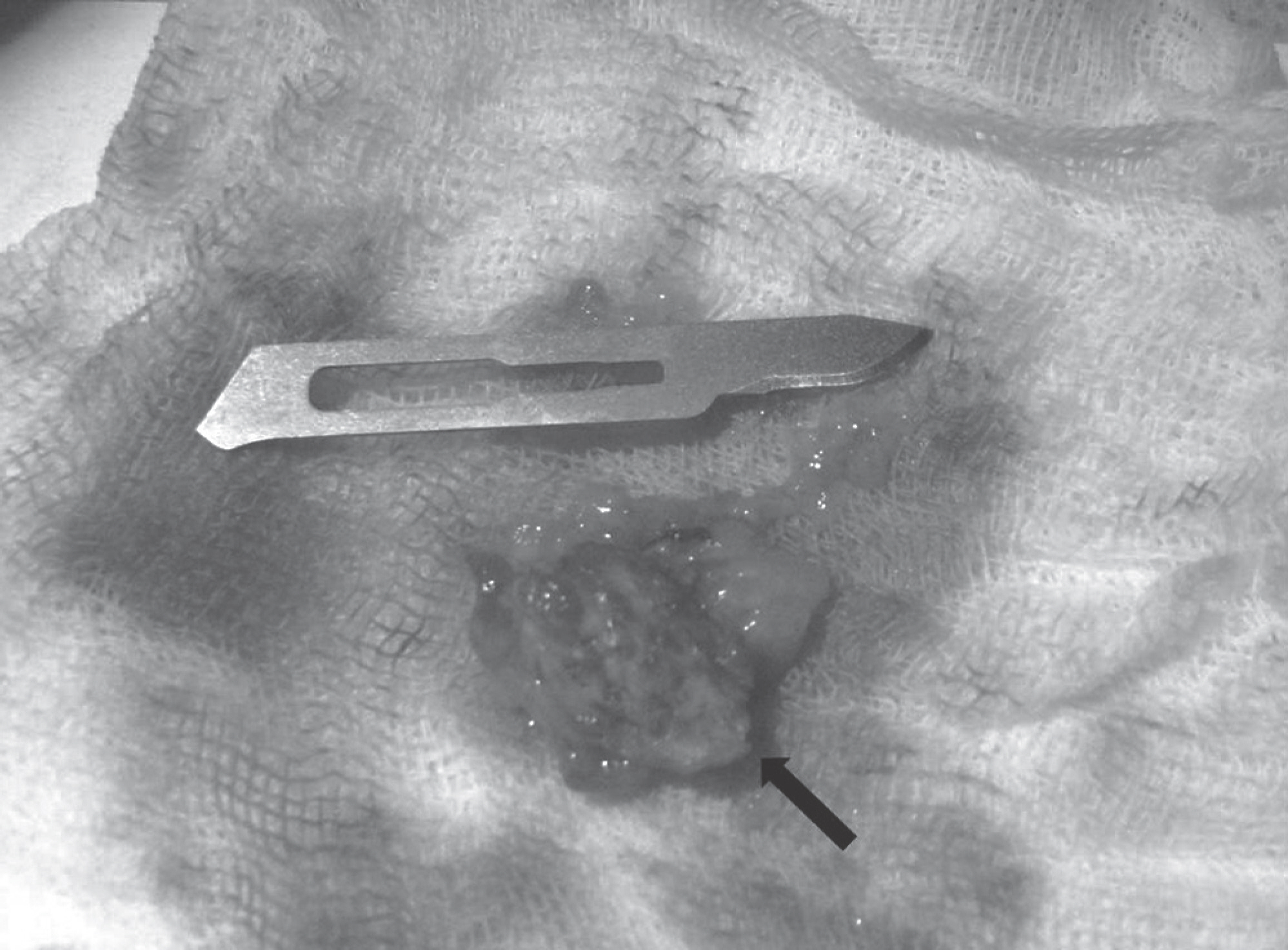
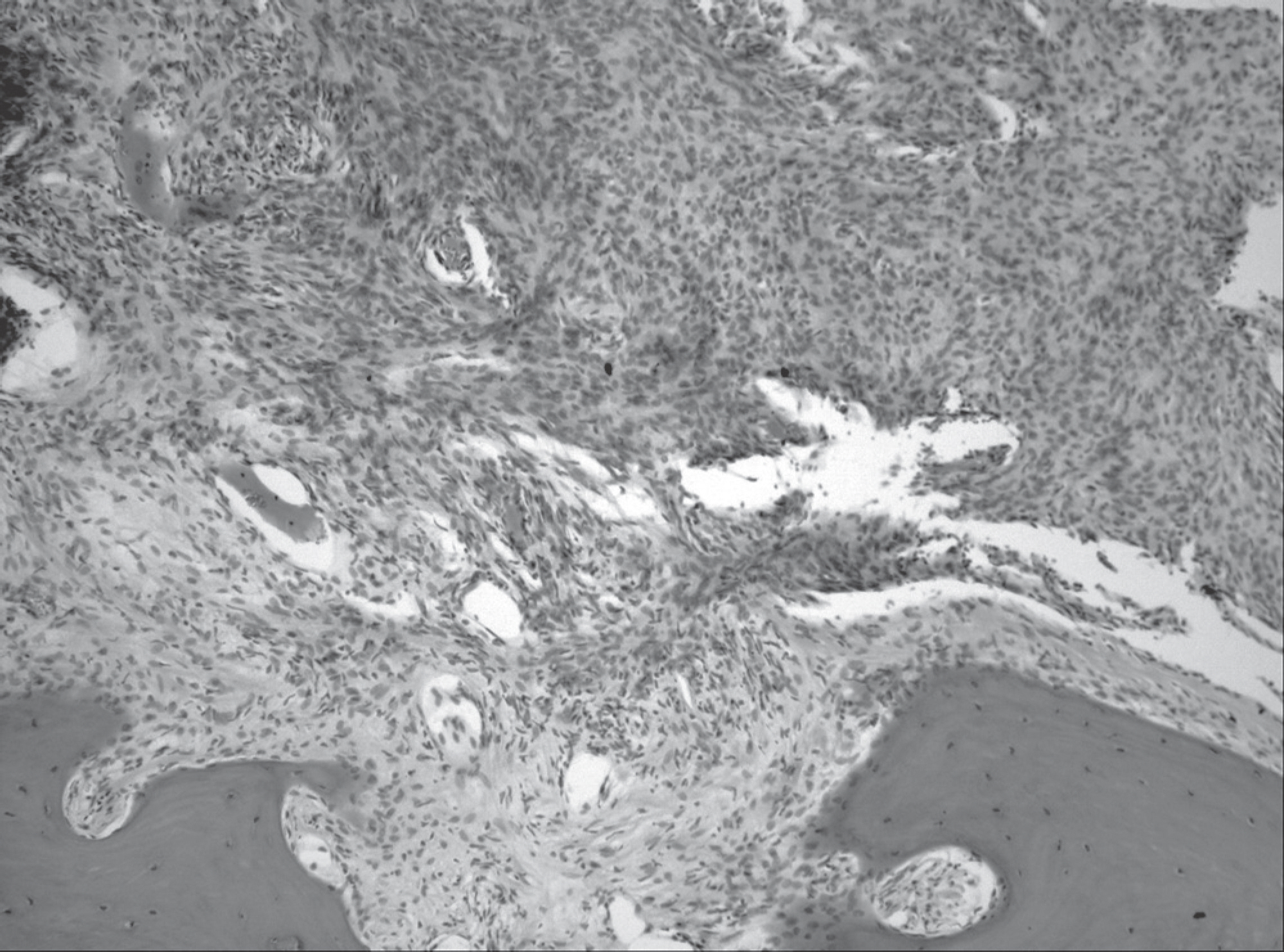
We experienced a 59-year-old patient who complained of weakness in the lower limbs and gait disturbance. Spinal cord compressing mass was discovered on a MRI at T6 and there was a vertical plate at the posterior side of the mass. Surgical finding showed complete ossification in the dural attachment site of the mass. Though the tumor mass could be excised with the inner layer of the dura mater en masse, more forceful retraction of the spinal cord was unavoidable than the other soft mass resection.
RESULTS
The preoperative neurological impairment improved after the surgery and she was able to walk well.
CONCLUSIONS
Ossification makes a resection difficult and vulnerable to develop neurological deterioration. But if we could suspect such an ossification through an image test, it would be helpful to make a surgical plan to avert a neurologic complication.
Keyword
Figure
Reference
-
1.Naderi S., Yilmaz M., Canda T., Acar U. Ossified thoracic spinal meningioma in childhood: a case report and review of the literature. Clin Neurol Neurosurg. 2001. 103:247–9.2.Doita M., Harada T., Nishida K., Marui T., Kurosaka M., Yoshiya S. Recurrent calcified spinal meningioma detected by plain radiograph. Spine (Phila Pa 1976). 2001. 26:E249–52.
Article3.Niijima K., Huang YP., Malis LI., Sachdev VP. Ossified spinal meningioma en plaque. Spine (Phila Pa 1976). 1993. 18:2340–3.
Article4.Saito T., Arizono T., Maeda T., Terada K., Iwamoto Y. A novel technique for surgical resection of spinal meningioma. Spine (Phila Pa 1976). 2001. 26:1805–8.
Article5.Rogers L. A spinal meningioma containing bone. J Br Surg. 1928. 15:675–7.
Article6.Freidberg SR. Removal of an ossified ventral thoracic meningioma. Case report. J Neurosurg. 1972. 37:728–30.7.Levy WJ Jr., Bay J., Dohn D. Spinal cord meningioma. J Neurosurg. 1982. 57:804–12.
Article8.Roux FX., Nataf F., Pinaudeau M., Borne G., Devaux B., Meder JF. Intraspinal meningiomas: review of 54 cases with discussion of poor prognosis factors and modern therapeu-tic management. Surg Neurol. 1996. 46:458–64.
Article9.Kubota T., Sato K., Yamamoto S., Hirano A. Ultrastructural study of the formation of psammoma bodies in fibroblastic meningioma. J Neurosurg. 1984. 60:512–7.
Article10.Huang TY., Kochi M., Kuratsu J., Ushio Y. Intraspinal os-teogenic meningioma: report of a case. J Formos Med As-soc. 1999. 98:218–21.


