Primary Yolk Sac Tumor of the Omentum: Case Report
- Affiliations
-
- 1Department of Radiology, Research Institute of Radiological Science, Yonsei University College of Medicine, Seoul, Korea. oytaik@yuhs.ac
- 2Department of Pathology, Yonsei University College of Medicine, Seoul, Korea.
- KMID: 2208872
- DOI: http://doi.org/10.3348/jksr.2012.66.1.57
Abstract
- A 32-year-old woman had been referred to our hospital for lower abdominal pain. Pelvic ultrasonography and magnetic resonance imaging revealed a huge solid mass with an internal cystic portion. The patient underwent a staging laparotomy and subsequent total abdominal hysterectomy with bilateral salpingo-oophorectomy, bilateral pelvic lymph nodes sampling, and total omentectomy. At staging laparotomy, a large omental mass was found. The tumor displayed the typical histological patterns observed in the yolk sac tumor. The alpha-fetoprotein (AFP) serum value on the 10th day after surgery was 11,576.67 IU/mL and decreased to 6.46 IU/mL after chemotherapy. At the end of the treatment, all the findings, including the AFP level, were normal. We report a case of primary yolk sac tumor of the omentum in a 32-year-old woman.
MeSH Terms
Figure
-
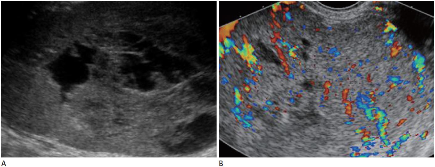
Fig. 1 Ultrasonography of the omental yolk sac tumor in a 32-year-old woman. A. Pelvic ultrasonography reveals a huge heterogeneous echogenic solid mass with internal anechoic portion. B. Color Doppler ultrasound shows internal hypervascularity of the tumor.
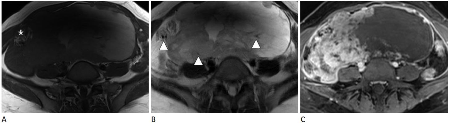
Fig. 2 Magnetic resonance imaging (MRI) of the omental yolk sac tumor in a 32-year-old woman. A. Axial T1-weighted MR image shows a multi-lobulated solid and cystic pelvic mass with some areas of hemorrhage (asterisk). B. Axial T2-weighted MR image shows multi-lobulated solid and cystic mass with internal signal voids (arrowheads) within the tumor due to a rich vascular supply. C. Axial postcontrast MR image shows a striking enhancement of the mass except for the area of hemorrhage and cysts.

Fig. 3 Positron emission tomography computed tomography (PET-CT) of the omental yolk sac tumor in a 32-year-old woman. A. Post-contrast CT scan shows an enhancing multi-lobulated solid mass with an internal cystic and necrotic portion. B. PET shows intense FDG uptake in the solid portion of the tumor, which suggests a malignant tumor. Note.-FDG = fludeoxyglucose
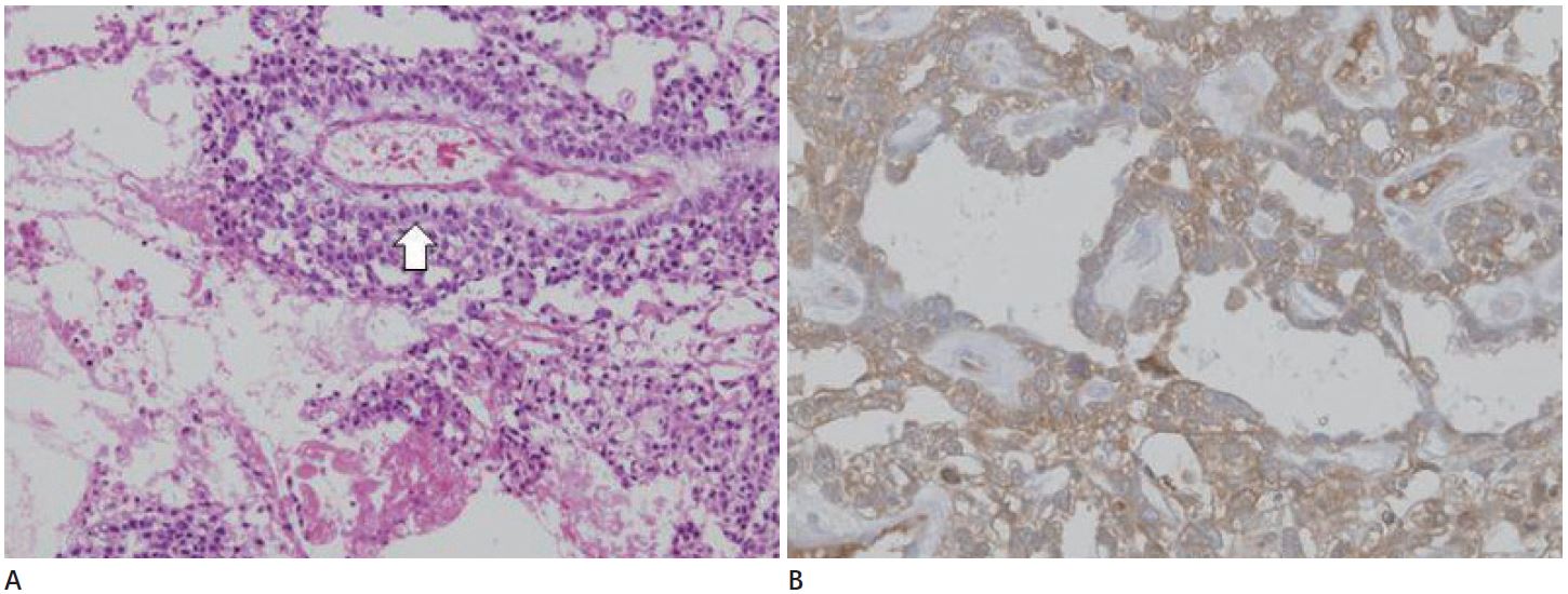
Fig. 4 Histological feature of the omental yolk sac tumor in a 32-year-old woman. A. Histological evaluation of the specimen exhibits typical Schiller-Duval body (arrow, H&E × 100). B. Immunohistochemical staining shows a positive reaction with alpha-fetoprotein (× 100).
Reference
-
1. Kim SW, Park JH, Lim MC, Park JY, Yoo CW, Park SY. Primary yolk sac tumor of the omentum: a case report and review of the literature. Arch Gynecol Obstet. 2009; 279:189–192.2. Clement PB, Young RH, Scully RE. Extraovarian pelvic yolk sac tumors. Cancer. 1988; 62:620–626.3. Gooneratne S, Keh P, Sreekanth S, Recant W, Talerman A. Anterior mediastinal endodermal sinus (yolk sac) tumor in a female infant. Cancer. 1985; 56:1430–1433.4. Dede M, Pabuccu R, Yagci G, Yenen MC, Goktolga U, Gunhan O. Extragonadal yolk sac tumor in pelvic localization. A case report and literature review. Gynecol Oncol. 2004; 92:989–991.5. Sompayrac SW, Mindelzun RE, Silverman PM, Sze R. The greater omentum. AJR Am J Roentgenol. 1997; 168:683–687.6. Oken MM, Creech RH, Tormey DC, Horton J, Davis TE, McFadden ET, et al. Toxicity and response criteria of the Eastern Cooperative Oncology Group. Am J Clin Oncol. 1982; 5:649–655.7. Jones MA, Clement PB, Young RH. Primary yolk sac tumors of the mesentery. A report of two cases. Am J Clin Pathol. 1994; 101:42–47.8. Yamaoka T, Togashi K, Koyama T, Ueda H, Nakai A, Fujii S, et al. Yolk sac tumor of the ovary: radiologic-pathologic correlation in four cases. J Comput Assist Tomogr. 2000; 24:605–609.9. Choi HJ, Moon MH, Kim SH, Cho JY, Jung DC, Hong SR. Yolk sac tumor of the ovary: CT findings. Abdom Imaging. 2008; 33:736–739.10. Xinghui Y, Jing H, Mingju L, Weizhong G. Endodermal sinus tumour of the omentum in a child. Pediatr Radiol. 2004; 34:985–987.
