Obstet Gynecol Sci.
2013 Nov;56(6):412-415. 10.5468/ogs.2013.56.6.412.
Primary omental yolk sac tumor
- Affiliations
-
- 1Department of Obstetrics and Gynecology, Yonsei University College of Medicine, Seoul, Korea. shkim70@yuhs.ac
- 2Department of Pathology, Yonsei University College of Medicine, Seoul, Korea.
- KMID: 1500377
- DOI: http://doi.org/10.5468/ogs.2013.56.6.412
Abstract
- Extra-ovarian yolk sac tumor arising in the omentum is extremely rare. As yolk sac tumor originated from the omentum has been rarely reported, its clinical information is very limited. The authors encountered a case of yolk sac tumor originated from the omentum, and reported the case herein. A 32-year-old woman was presented with developed low abdominal distension for a month. Magnetic resonance imaging findings were suggestive of ovarian malignancy with ascites and peritoneal seeding nodules. Explorative laparotomy was performed and then the findings from frozen biopsy of omentum were suggestive of poorly differentiated tumor though whether it was primary or metastatic was uncertain. Thus, staging laparotomy were performed. Histopathology confirmed that the tumor was a yolk sac tumor of omentum origin. Then, 6 cycles of postoperative adjuvant chemotherapy at intervals of 3 weeks were performed using bleomycin, etoposide, and cisplatin regimen. Four-year outpatient follow-up thereafter showed no relapse.
MeSH Terms
Figure
-
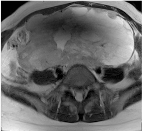
Fig. 1 Magnetic resonance imaging (T2) finding. About 8×16×18 cm sized mass is seen on pelvis. Ascites and peritoneal seeding nodules are suggested.

Fig. 2 Histological examination by H&E. Schiller-Duval body. The glomerular-like process exists in the empty space, and surrounded by tumor cells. The process has blood vessels in its center (A, ×100; B, ×200). Immunohistochemistry for alpha-fetoprotein (AFP). It revealed a cytoplasmic expression of AFP in a subset of tumor cells (C, ×40).
Reference
-
1. Pasternack T, Shaco-Levy R, Wiznitzer A, Piura B. Extraovarian pelvic yolk sac tumor: case report and review of published work. J Obstet Gynaecol Res. 2008; 34:739–744.2. Moller H, Evans H. Epidemiology of gonadal germ cell cancer in males and females. APMIS. 2003; 111:43–46.3. Oken MM, Creech RH, Tormey DC, Horton J, Davis TE, McFadden ET, et al. Toxicity and response criteria of the Eastern Cooperative Oncology Group. Am J Clin Oncol. 1982; 5:649–655.4. Kim SW, Park JH, Lim MC, Park JY, Yoo CW, Park SY. Primary yolk sac tumor of the omentum: a case report and review of the literature. Arch Gynecol Obstet. 2009; 279:189–192.5. Jones MA, Clement PB, Young RH. Primary yolk sac tumors of the mesentery. A report of two cases. Am J Clin Pathol. 1994; 101:42–47.6. Talerman A, Haije WG, Baggerman L. Serum alphafetoprotein (AFP) in diagnosis and management of endodermal sinus (yolk sac) tumor and mixed germ cell tumor of the ovary. Cancer. 1978; 41:272–278.7. Kommoss F, Schmidt M, Merz E, Knapstein PG, Young RH, Scully RE. Ovarian endometrioid-like yolk sac tumor treated by surgery alone, with recurrence at 12 years. Gynecol Oncol. 1999; 72:421–424.8. Williams S, Blessing JA, Liao SY, Ball H, Hanjani P. Adjuvant therapy of ovarian germ cell tumors with cisplatin, etoposide, and bleomycin: a trial of the Gynecologic Oncology Group. J Clin Oncol. 1994; 12:701–706.
